A complete listing of currently available online programs is provided below. To view course materials click an available viewing format provided with each listing (PDF, HTML, Interactive). To access online exams and claim credit you must be registered and logged in. To add courses to your "MyAR Archives" user account select the "Add To Cart" button provided with each course title and follow the prompts.
|
* Pediatric MRI with Gadopiclenol
|
|
Released:
October 16, 2024
•Expires:
October 31, 2027
|
CE credits:
1.0
• Cost:
$0.00
|
|
Faculty:
Azam Eghbal, MD
|
While gadolinium-based contrast agents (GBCAs) are essential to performing MR imaging, guidance from the US Food and Drug Administration (FDA) and the American College of Radiology (ACR) includes the recommendation of limiting a patient's gadolinium exposure. One technique for reducing gadolinium exposure is to use a high- relaxivity gadolinium-based contrast agent (GBCA) that can be administered at a lower dose than a conventional GBCA without deteriorating image quality. Gadopiclenol recently approved by the FDA is a high-relaxivity GBCA, designed to reveal high-quality images at half the conventional dose of other GBCAs.
This educational program provides the clinical rationale for adopting the use of Gadopiclenol for pediatric MRI.
Through case study reviews, Azam Eghbal, MD, will share clinical insights to achieve optimal image quality using a lower administered gadolinium dose when performing pediatric MRI. A focus on the selection and use of a high relaxivity, lower dose GBCA in vulnerable patients, receiving multiple MR contrast injections, will be supported by clinical evidence and imaging protocols. Audience participation will be encouraged during the Q & A session following the presentation.
Educational Objectives:
At the completion of this activity, participants should be able to:
-
Explain the properties that distinguish gadopiclenol from other GBCAs.
-
Apply clinical considerations for lower gadolinium dosing options using gadopiclenol for pediatric MRI.
-
Implement gadopiclenol MR imaging protocols to achieve optimal imaging and diagnostic confidence in pediatric MRI.
-
Employ the use of gadopiclenol in vulnerable patient populations who may experience repeat, lifetime dosing and scanning.
This program is an educational activity sponsored by Guerbet, LLC.
|
|
|
|
|
* Neuro MRI with Gadopiclenol
|
|
Released:
October 8, 2024
•Expires:
October 31, 2027
|
CE credits:
1.0
• Cost:
$0.00
|
|
Faculty:
Pareen Mehta, MD
|
While GBCAs are essential to performing neuro MR imaging, guidance from the US Food and Drug Administration (FDA) and the American College of Radiology (ACR) includes the recommendation of limiting a patient's gadolinium exposure
One technique for reducing gadolinium exposure is to use a high-relaxivity gadolinium-based contrast agent (GBCA) that can be administered at a lower dose than a conventional GBCA without deteriorating image quality. Gadopiclenol, recently received FDA approval and is a high-relaxivity GBCA, designed to generate high-quality images at half the conventional dose of other GBCAs
This educational program provides the clinical rationale for adopting the use of Gadopiclenol for neuro MRI and includes practice considerations for changing from one GBCA to another.
Through case study reviews, Pareen Mehta, MD, will share clinical insights into achieving optimal image quality using a lower administered gadolinium dose when performing neuro MRI.
Educational Objectives:
At the completion of this activity, participants should be able to:
-
Understand and employ the clinical indications for neuro MRI using gadopiclenol.
-
Apply clinical considerations for lower neuro MRI gadolinium dosing protocols as compared to other GBCAs in neuroradiology.
-
Utilize neuro MR imaging protocols using gadopiclenol to achieve optimal image quality and diagnostic confidence.
This program is an educational activity sponsored by Guerbet, LLC.
|
|
|
|
|
* Breast MRI with Gadopiclenol
|
|
Released:
July 18, 2024
•Expires:
July 31, 2027
|
CE credits:
1.0
• Cost:
$0.00
|
|
Faculty:
Katja Pinker-Domenig, MD, PhD, EBBI, FISMRM, FSBI
|

Dynamic contrast-enhanced breast MR imaging using gadolinium-based contrast agents (GBCAs) offers superior cancer detection to mammography and breast ultrasound due to the visualization of tumor neovascularity as a tumor-specific feature. Despite the clear advantages of breast MR imaging, it is currently recommended only for patients with a high lifetime risk of breast cancer, but recent data shows that there is also a benefit for women at a greater than average risk
While GBCAs are essential to performing breast MR imaging, guidance from the US Food and Drug Administration (FDA) and the American College of Radiology (ACR) includes the recommendation of limiting a patient's gadolinium exposure where possible. One technique for reducing gadolinium exposure is to use a high-relaxivity GBCA that can be administered at a lower dose than a conventional GBCA without deteriorating image quality. Gadopiclenol recently received FDA approval and is a high-relaxivity GBCA that decreases gadolinium exposure.
This educational program provides a clinical rationale for lowering the administered gadolinium dose when performing breast MRI on patients who may receive repeat MR contrasted exams throughout their lifetime for breast cancer screening. Through a review of case studies, Dr. Pinker-Domenig will provide insight on how to achieve optimal imaging while lowering the gadolinium dose with Gadopiclenol for breast MRI.
Educational Objectives:
At the completion of this activity, participants should be able to:
-
Understand and employ the clinical indications for dynamic-contrast enhanced breast MRI.
-
Access clinical considerations and apply lower gadolinium dosing options for patients who may experience repeat, lifetime dosing of a GBCA for breast MRI.
-
Implement breast MR imaging protocols to achieve the most accurate breast cancer diagnosis.
This program is an educational activity sponsored by Guerbet, LLC.
|
|
|
|
|
* Advanced MRI Safety Training For Healthcare Professionals (4th Edition): Level 2 MR Personnel
|
|
Released:
April 22, 2020
•Expires:
March 31, 2025
|
CE credits:
3.0
• Cost:
$50.00
|
|
Faculty:
Frank G. Shellock, PhD, FACC, FACR, FACSM
|
This program reviews fundamental MRI safety information and meets the annual training recommendations from the American College of Radiology. Importantly, MRI facilities must now comply with the revised requirements for diagnostic imaging from The Joint Commission and document that MRI technologists participate in ongoing education that includes annual training on safe MRI practices in the MRI environment. Notably, Advanced MRI Safety Training for Healthcare Professionals, Level 2 MR Personnel covers each MRI safety topic specified by The Joint Commission, as well as many additional subjects that will expand the knowledge-base of healthcare professionals involved with MRI technology.
With 35 years of experience in the field of MRI, the author of the best-selling textbook series, the Reference Manual for Magnetic Resonance Safety, Implants and Devices, and the creator of the internationally popular website, MRIsafety.com, Dr. Frank G. Shellock is uniquely qualified to present the information in this program.
Advanced MRI Safety Training for Healthcare Professionals (4th Edition), Level 2 MR Personnel is a 2 hour and 45 minute program that is divided into three different sections.
Educational Objectives
-
Understand the safety issues related to MRI.
-
Describe the bioeffects associated with the static magnetic field, time-varying magnetic fields, and radiofrequency fields.
-
Present guidelines that prevent projectile-related accidents.
-
Describe polices that avoid issues related to acoustic noise.
-
Review procedures that prevent burns associated with MRI.
-
Explain and demonstrate appropriate pre-MRI screening procedures.
-
Identify techniques to manage patients with claustrophobia, anxiety, or emotional distress.
-
Describe guidelines to handle medical emergencies in the MRI setting.
-
Understand the safety considerations for gadolinium-based contrast agents.
This is a Pay-To-View program. Purchase is required for full program access.
|
|
|
|
|
* Basic MRI Safety Training (4th Edition): Level 1 MR Personnel
|
|
Released:
May 3, 2023
•Expires:
May 31, 2026
|
CE credits:
1.0
• Cost:
$30.00
|
|
Faculty:
Frank G. Shellock, PhD, FACC, FACR, FACSM
|
Individuals entering the magnetic resonance imaging (MRI) environment, whether on a regular or infrequent basis, must be properly trained to ensure their safety, the protection of patients, and the security of other facility staff members. This program, Basic MRI Safety Training, Level 1 MR Personnel accomplishes the initial training that is necessary to ensure safety in the unique setting associated with the MRI system. It includes information pertaining to MRI technology, describes common hazards and unique dangers associated with the MRI setting, and presents vital recommendations and guidelines to prevent accidents and injuries. Importantly, this program provides the fundamental MRI safety information for Level I MR Personnel recommended by the American College of Radiology and may be utilized by individuals preparing for safety training as Level 2 MR Personnel.
With more than 35 years of experience in the field of MRI, the author of the best-selling textbook series, the Reference Manual for Magnetic Resonance Safety, Implants and Devices, and the creator of the internationally popular website, www.MRIsafety.com, Dr. Frank G. Shellock is uniquely qualified to present the information in this program.
Educational Objectives
-
Appreciate the importance of MRI
-
Identify the hazards associated with MRI
-
Understand the screening process
-
Describe steps to prevent accidents and injuries
This is a Pay-To-View program. Purchase is required for full program access.
|
|
|
|
|
* The Online Course for MR Safety Officers (MRSO) and MR Medical Directors (MRMD)
|
|
Released:
September 29, 2022
•Expires:
September 30, 2025
|
CE credits:
10.0
• Cost:
$750.00
|
|
Faculty:
William Faulkner, B.S.,R.T.(R)(MR)(CT), FSMRT | Kristan Harrington, MBA, R.T.(R)(MR), ARRT, MRSO
|
Applied Radiology and William Faulkner & Associates are pleased to introduce “The Online Course for MR Safety Officers (MRSO) and MR Medical Directors (MRMD)”. This comprehensive program, focusing on MR Safety, covers many aspects relating to the safety practices of the Magnetic Resonance Imaging (MRI) environment. It is designed for those individuals who are either currently serving as their facility’s MRSO and/or MRMD, or those who are preparing to assume these responsibilities. The content of this course will be helpful for those preparing for the American Board of Magnetic Resonance Safety MRSO and MRMD examinations.
The content of The Online Course for MR Safety Officers and MR Medical Directors is based upon current FDA and ACR guidelines, including the ACR Guidance Document for MR Safe Practices, as well as those promulgated by industry regulatory bodies such as the International Electrotechnical Commission.
The complete online program has been approved for up to 10 AMA PRA Category 1 credits™ (CME) and 10 Category A ARRT continuing educational credits (CE).
Module 1: Basic MRI Physics Relevant to MRI Safety
Module 2: Static Field: Bioeffects and Access Control
Module 3: Gradient Magnetic Fields: Bioeffects and Safety
Module 4: Radio Frequency Field: Bioeffects and Safety
Module 5: Implants and Devices
Module 6: Gadolinium-Based MR Contrast Agents
Module 7: MR Safety Screening
Module 8: Managing Emergent Situations and Patient Considerations: Quench and Patient Anxiety & Patient Monitoring
The Online Course for MR Safety Officers (MRSO) and MR Medical Directors (MRMD) is not affiliated with, nor endorsed by the American Board of Magnetic Resonance Safety (ABMRS)
Terms and Conditions of Use: Click To View
This is a Pay-To-View program. Purchase is required for full program access.
|
|
|
|
|
Advanced CT Technology: Improving Workflow and Cost of Ownership
|
|
Released:
January 19, 2023
•Expires:
January 31, 2026
|
CE credits:
1.5
• Cost:
$0.00
|
|
Faculty:
Lawrence N. Tanenbaum, MD, FACR | Rick Banner
|
CT Technology continues to evolve with modern platforms now being designed with improved patient comfort, increased workflow, and advanced applications supporting comprehensive diagnoses, precise treatment plans and better patient outcomes.
In this CE accredited Expert Forum Webinar, Rick Banner, RT (R), Senior Director of Marketing, Fujifilm Medical Systems U.S.A., Inc. shares insights into the design of advanced CT platforms that are addressing today’s clinical challenges while reducing the total cost of ownership. These advanced features will be supported by Dr. Lawrence N. Tanenbaum’s personal experience on how these advanced features are making a clinical difference in the day-to-day operations in busy imaging departments.
Educational Objectives:
At the completion of this activity, participants should be able to:
-
Implement advanced CT applications
-
Effectively manage obese and claustrophobic patients
-
Implement workflow considerations with improved CT service �and reliability
Supported through an educational grant from FUJIFILM Medical Systems U.S.A., Inc.
|
|
|
|
|
Cardiovascular Imaging: Complex Applications in Cardiac CT and CT Angiography
|
|
Released:
June 8, 2021
•Expires:
July 31, 2026
|
CE credits:
1.0
• Cost:
$0.00
|
|
Faculty:
Richard Hallett, MD
|
Often there is lack of knowledge amongst imaging professionals regarding IV contrast dynamics and the relationship between contrast injection, observed enhancement, and scan acquisition timing. The design of appropriate imaging protocols leads to effective and consistent cardiovascular exams; especially when applied to complex clinical scenarios.
This CME/CE accredited program will review contrast-saline dynamics and discuss rational protocol design for cardiac and vascular CT angiography. Effective cardiovascular imaging protocols, utilizing multiple CT injector platforms to achieve optimal imaging, will be reviewed. We invite you to join Dr. Richard Hallett for a comprehensive discussion and case study review of this important topic.
Following the presentation questions from the audience were addressed in a moderated Q&A session.
Educational Objectives:
At the completion of this activity, participants should be able to:
-
Understand the relationship between bolus contrast media injection and observed enhancement.
-
Implement methods to design rational contrast injection / scan acquisition protocols.
-
Review various models of CT contrast injectors and discuss benefits, limitations, and injection protocol adjustments for each.
-
Apply customized contrast-saline injection / scan principles to complex cardiovascular disease cases.
Made possible through an unrestricted educational grant from Bracco Diagnostics, Inc.
|
|
|
|
|
Clinical Applications & Imaging Considerations of Gadopiclenol Injection
|
|
Released:
May 10, 2023
•Expires:
May 31, 2026
|
CE credits:
1.0
• Cost:
$0.00
|
|
Faculty:
Lawrence N. Tanenbaum, MD, FACR
|
In September 2022, Gadopiclenol injection, which is both macrocyclic (stable) and has high relaxivity properties received FDA marketing approval and may provide dosing options that can decrease gadolinium exposure, particularly in vulnerable patients.
In this accredited Expert Forum webinar, the assessment of Gadopiclenol injection in neuro, body, and pediatric MR imaging will be presented by a panel of MR imaging experts and will include commentary from Lawrence N. Tanenbaum, MD. To encourage audience participation, a live Q & A session will follow the discussion.
Educational Objectives:
At the completion of this activity, participants should be able to:
-
Explain the properties that distinguish Gadopiclenol injection from the other high-relaxivity and conventional gadolinium-based contrast agents.
-
Describe the clinical use of Gadopiclenol injection in neuro, body, and pediatric MRI applications.
-
Describe the clinical development program of Gadopiclenol injection for neuro and body MRI applications.
-
Apply clinical considerations and dosing options when selecting a macrocyclic, high relaxivity, MRI contrast in vulnerable patient populations.
This program has been sponsored by Guerbet LLC.
|
|
|
|
|
Clinical Applications of MR Imaging in Stereotactic Localization Radiosurgery Procedures
|
|
Released:
January 2, 2024
•Expires:
January 31, 2027
|
CE credits:
1.5
• Cost:
$0.00
|
|
Faculty:
Jenny WU, MD | Glen Stevens, DO, PhD
|
Gadolinium-based contrast agents (GBCAs) are essential for enhancing the quality of MRI scans to make it easier for radiologists to identify pathology. The US Food and Drug Administration (FDA) and the American College of Radiology (ACR) recommend limiting patient exposure to gadolinium where possible.
One technique for reducing this exposure is to use a high relaxivity GBCA that can be administered at a lower dose than a conventional GBCA, without any impact on image quality. GBCA selection for interventional procedures such as radiosurgery is one such example, where the selection of a GBCA can play a role in patient management and outcomes.
In this program, an experienced neuroradiologist and neuro oncologist will present their clinical experiences with the use of stereotactic localization radiosurgery, following the administration of a high relaxivity GBCA. Through case study examples, the clinical impact of contrast selection will be demonstrated. Following the presentation, questions from the audience will be addressed during a live Q&A.
Educational Objectives:
At the completion of this activity, participants should be able to:
-
Review the role of stereotactic radiosurgery (SRS) for brain metastases and the role of pre-operative localization MRI exams for stereotactic radiosurgery.
-
Outline the differences between gadopiclenol and other group II gadolinium-based contrast agents.
-
Discuss the use of gadopiclenol in specific case-based scenarios for stereotactic localization exams and its impact on patient management.
This program is made possible through an unrestricted educational grant from Guerbet.
|
|
|
|
|
Clinical Utility of Barium Sulfate Products: Formulation Determines Appropriate Use
|
|
Released:
June 14, 2021
•Expires:
July 31, 2026
|
CE credits:
0.75
• Cost:
$0.00
|
|
Faculty:
Applied Radiology
|
Steven Sireci, MD
Barium-containing contrast media contain barium sulfate (BaSO4), as well as a number of additives. The BaSO4 provides radiodensity (ie, radio-opacity) and physical density (g/mL) to the suspension. Depending on the intended use of the contrast, the specific combination of additives provide other useful properties, including: BaSO4 particle suspension; mucosal coating; viscosity and texture; sweetness; filling and laxative effects; preservation; foaming prevention; and flavoring. Here we describe the purpose of each ingredient and additive found in BaSO4 contrast media products currently available for fluoroscopic and radiographic examinations. In addition, we review the role of these products in anatomic radiographic evaluation of the gastrointestinal (GI) tract and in evaluation of the physiology of swallowing. Finally, we review barium preparations used in computed tomography (CT) applications.
At the conclusion of this activity, participants will be able to:
-
Explain the role that BaSO4 plays in barium-containing contrast media;
-
Review the purpose of each ingredient/additive found in BaSO4 contrast media products available for fluoroscopic and radiographic examinations;
-
Describe the formulation of barium products that are appropriate for single- and double-contrast anatomic radiographic evaluation of the gastrointestinal (GI) tract;
-
Summarize qualities of the barium-containing contrast media used in evaluation of swallowing that make them most appropriate for that application; and,
-
State the properties of barium-containing contrast media used in computed tomography (CT) applications.
This program has been supported through an educational grant from Bracco Diagnostics, Inc.
|
|
|
|
|
Clinical Utility of Barium Sulfate Products: Formulation Determines Appropriate Use
|
|
Released:
November 24, 2021
•Expires:
April 30, 2027
|
CE credits:
1.0
• Cost:
$0.00
|
|
Faculty:
Steven Sireci, MD
|
Depending on its intended application in Fluoroscopy or CT, Barium Sulfate (BaSO4) contains specific combinations of additional ingredients that support the diagnosis of disease, define treatment options, and improve patient outcomes. When used for the anatomical evaluation of the GI tract or the physiology of swallowing, a comprehensive understanding of the clinical utility of Barium products and their additives, is needed. This accredited program explains the differences and the benefits of each additive and is designed to increase knowledge about the clinical applications of Barium.
Dr. Sireci will provide a comprehensive overview of each Barium product, its indication and intended use. Following the presentation, the audience is invited to join Dr. Sireci for lively Q & A session.
Educational Objectives:
At the completion of this activity, participants should be able to:
-
Explain the role that BaSO4 plays in barium-containing contrast media.
-
Review the purpose of each ingredient/additive found in BaSO4 contrast media products currently available for fluoroscopic and radiographic examinations.
-
Describe the formulation of barium products that are appropriate for single and double contrast anatomic radiographic evaluation of the gastrointestinal (GI) tract.
-
Summarize qualities of the barium-containing contrast media used in evaluation of swallowing that make them most appropriate for that application.
-
State the properties of barium-containing contrast media used in computed tomography (CT) applications
Educational Credits
One (1) AHRA CRA Credit
Made possible through an educational grant from Bracco Diagnostics, Inc.
|
|
|
|
|
Contrast Media Basics: Important Considerations for the Pharmacist
|
|
Released:
March 1, 2024
•Expires:
March 31, 2027
|
CE credits:
2.5
• Cost:
$0.00
|
|
Faculty:
William Faulkner, B.S.,R.T.(R)(MR)(CT), FSMRT | Kristan Harrington, MBA, R.T.(R)(MR), ARRT, MRSO | Rutu Patel, PharmD, RPh
|
Applied Radiology has developed this two-part, on-demand webinar and accompanying digital monograph to increase overall understanding of the properties, clinical benefits, and safety considerations of iodinated and gadolinium-based contrast agents (GBCAs). The goal of this program is to fully prepare pharmacy personnel to make informed decisions regarding contrast media selection.
The complete online program has been approved for up to 2.5 ACPE and Category A ARRT continuing educational credits.
Part 1: Contrast Media Basics
Part 2: A Pharmacist’s Guide to Gadolinium-Based Contrast Agents
Part 3: Digital Monograph: Contrast Media Basics: A Comprehensive Overview
This program was supported through an educational grant from Bracco Diagnostics, Inc.
|
|
|
|
|
Contrast-Enhanced MRI: Optimizing Imaging of the Brain and Spine
|
|
Released:
November 24, 2021
•Expires:
December 31, 2026
|
CE credits:
1.0
• Cost:
$0.00
|
|
Faculty:
Lawrence N. Tanenbaum, MD, FACR | Susie Bash, MD
|
In this Expert Forum, Lawrence Tanenbaum, MD, FACR and Suzie Bash, MD review considerations for optimizing contrast-enhanced MRI in neuroimaging, extending from patient tolerance to standardizing procedures. Following the presentation questions from the audience were addressed in a moderated Q&A session.
Educational Objectives:
-
Manage issues related to patient tolerability and acute adverse events.
-
Implement standardized and optimized MR contrast-enhanced protocols.
-
Identify clinical indications for contrast-enhanced MRI for brain and spine imaging
Supported by GE Healthcare
|
|
|
|
|
Development and Use of Elucirem™ (gadopiclenol) Injection, a Macrocyclic, High-relaxivity GBCA for CE-MRI
|
|
Released:
February 24, 2023
•Expires:
March 31, 2026
|
CE credits:
1.0
• Cost:
$0.00
|
|
Faculty:
Applied Radiology
|
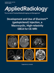
After decades of administrations, substantial evidence has accumulated for the efficacy and safety of gadolinium-based contrast agents (GBCAs) in magnetic resonance imaging (MRI). However, their excellent performance has led to increased use and dosing for many neuro and cardiovascular applications in adults and children.
As a result, concern has been growing in the radiology community, particularly with respect to use of the less stable agents, which are generally linear and, therefore, bind less tightly to the gadolinium (Gd) ion. While recognizing the value of these agents, the US Food and Drug Administration (FDA) and the American College of Radiology (ACR) now recommend limiting patient Gd exposure where possible.
One technique for reducing Gd exposure is to use a high-relaxivity GBCA that can be used at a lower dose than a conventional GBCA without impact on image quality. Until recently, however, the GBCA with the highest relaxivity was a linear agent. Consequently, it has been widely recognized for some time that it would be preferable if a GBCA was both macrocyclic in structure and high relaxivity, and that such an agent could be used to reduce Gd exposure by decreasing the amount of Gd injected.
A new GBCA that is both macrocyclic and high relaxivity has been developed and received FDA marketing approval in September 2022. A group of radiologists with extensive experience in contrast-enhanced (CE)-MRI convened to discuss this new GBCA, Elucirem™ (gadopiclenol) injection, and this monograph presents a summary of this expert panel forum.
At the conclusion of this activity, participants will be able to:
-
Describe the properties that distinguish Elucirem from the other high-relaxivity and conventional GBCAs;
-
Demonstrate an understanding of the clinical development program of Elucirem for neuro and body imaging;
-
Define the patient populations that are most vulnerable to higher and/or repeat dosing of GBCAs; and,
-
Explain the factors to be considered when deciding to use MRI contrast in vulnerable patient populations.
This content is supported by Guerbet, LLC.
|
|
|
|
|
Diagnosis and Management of Swallowing Physiology: Standardized Contrast, the MBSImP™, & the IDDSI Framework
|
|
Released:
May 3, 2023
•Expires:
May 31, 2026
|
CE credits:
1.0
• Cost:
$0.00
|
|
Faculty:
Applied Radiology
|
Catriona M. Steele, PhD, Bonnie Martin-Harris, PhD, Memorie Gosa, PhD, Stephanie Edwards Allen, BSRT, MBA
The MBSS, also known as a video fluoroscopic swallow study (VFSS), is a barium-sulfate–enhanced fluoroscopic motion study used to evaluate anatomy and swallowing physiology simultaneously in real time. Varibar, an FDA-approved barium sulfate product for evaluation of dysphagia, together with The Modified Barium Swallow Impairment Profile (MBSImP), are used to ensure standardization and reproducibility in the MBSS. This monograph explains the relationship between Varibar, the MBSImP, and the International Dysphagia Diet Standardisation (IDDSI) framework in evaluation of swallowing in clinical practice.
At the conclusion of this activity, participants will be able to:
-
Explain how the modified barium swallow study (MBSS), The Modified Barium Swallow Impairment Profile (MBSImP), and barium sulfate products are used to evaluate dysphagia
-
Describe the development of FDA-approved barium sulfate products used to assess dysphagia
-
Review the International Dysphagia Diet Standardisation (IDDSI) framework: what it is and how it was developed
-
Define the relationship between the MBSS, the MBSImP, barium sulfate, and IDDSI, and how they are used in clinical practice
-
Summarize pediatric and clinical-practice considerations in performing the MBSS
This program has been supported through an educational grant from Bracco Diagnostics, Inc.
|
|
|
|
|
Evidence-based Approach to Minimizing IV Contrast Extravasations
|
|
Released:
July 19, 2019
•Expires:
January 31, 2027
|
CE credits:
1.0
• Cost:
$0.00
|
|
Faculty:
Ryan K Lee, MD, MBA
|
IV contrast extravasation is a not uncommon complication in the performance IV contrast enhanced CT examinations. In this accredited Expert Forum Webcast, we will review the different complications related to IV contrast administration. This will be followed by analysis of different factors that contribute to IV contrast-extravasation and subsequent discussion of best practices that can reduce the incidence of IV contrast extravasation. A novel method which reduced contrast extravasation by over 50% will also be presented.
Following the presentation questions from the audience were addressed in a moderated Q&A session.
Learning Objectives
-
Discuss the different types of complications associated with IV contrast extravasation.
-
Describe the various factors that can contribute to IV contrast extravasation.
-
Apply best practices to reduce the incidence of IV contrast extravasation.
No special educational preparation is required for this CME/CE Activity!
Supported through an unrestricted educational grant from Bracco Diagnostics, Inc.
|
|
|
|
|
Fluoroscopic Evaluation of the Bariatric Surgery Patient
|
|
Released:
November 1, 2020
•Expires:
December 31, 2026
|
CE credits:
1.0
• Cost:
$0.00
|
|
Faculty:
Applied Radiology
|
Cheri Canon, MD, and Jayleen Grams, MD, PhD
Obesity has been linked to increases in several clinically significant comorbidities. To address these related health issues, the number of bariatric procedures is growing. This monograph describes the role Fluoroscopy plays in the evaluation of the bariatric patient including a review of the imaging protocols and their necessary findings; designed to guide clinical decisions regarding which bariatric procedure is most appropriate for the patient.
At the conclusion of this activity, participants will be able to:
-
Review the incidence of obesity and the role of bariatric surgery as a strategy to address it
-
Explain the advantages of fluoroscopy in the evaluation of the bariatric patient
-
Summarize the considerations when performing fluoroscopic evaluation of the pre- and post-operative bariatric patient
-
Describe the protocols for fluoroscopic imaging of the gastrointestinal (GI) tract, including single- and dual-phase esophagrams and upper GI examinations
-
Detail the main challenges to performing fluoroscopy in bariatric patients
-
Relate the value of communication between the radiologist and bariatric surgeon
This Program Has Been Supported Through An Unrestricted Educational Grant from Bracco Diagnostics, Inc.
|
|
|
|
|
Gadopiclenol | Considerations for Use in Pediatric MRI
|
|
Released:
April 29, 2024
•Expires:
April 30, 2027
|
CE credits:
1.0
• Cost:
$0.00
|
|
Faculty:
Shreyas Vasanawala, MD, PhD
|
This educational program featuring Dr. Shreyas Vasanawala will cover pediatric MR imaging applications using Gadopiclenol, a recently approved gadolinium-based contrast agent (GBCAs).
Following a brief review of the literature and the currently available GBCAs, Dr. Vasanawala will discuss practice considerations as they relate to MRI contrast media selection and dose administration. Additionally, Dr. Vasanawala will share several case studies demonstrating the clinical value and benefits of Gadopiclenol for patients who may present with complex pathology and/or who may be subject to repeat contrast-enhanced MRI exams over their lifetime.
Educational Objectives:
At the completion of this activity, participants should be able to:
-
Explain the properties that distinguish gadopiclenol injection from the other high relaxivity gadolinium-based contrast agents (GBCAs).
-
Employ the FDA-approved indications and clinical applications for gadopiclenol injection in pediatric patients.
-
Apply clinical considerations and dosing options for gadopiclenol administered to vulnerable patient populations who may experience repeat, lifetime dosing of a GBCA.
This program is made possible through an unrestricted educational grant from Bracco Diagnostics Inc.
|
|
|
|
|
Gadopiclenol Injection | Practical Considerations for Use
|
|
Released:
October 23, 2023
•Expires:
October 31, 2026
|
CE credits:
1.0
• Cost:
$0.00
|
|
Faculty:
William Faulkner, B.S.,R.T.(R)(MR)(CT), FSMRT
|
With the recent FDA approval of gadopiclenol injection, radiologists now have the choice of a macrocyclic gadolinium-based contrast agent (GBCA) with 2 – 3 times higher relaxivity at a lower standard dose as compared to other gadolinium-based contrast agents on the marke
Therefore, and because gadopiclenlol is different than other currently available GBCAs, a review of the clinical benefits of using a higher relaxivity GBCA at a lower administered dose is important. A clear understanding of relaxivity and calculation of the volume to be administered based on dosing considerations, is necessary
In this accredited webinar, Mr. Faulkner will provide insights into the differences of the current FDA-approved GBCAs and how these differences apply to MR imaging. Following the presentation, questions from the audience were addressed during a live Q&A.
Educational Objectives:
At the completion of this activity, participants should be able to:
-
Identify the properties that distinguish gadopiclenol injection from the other high relaxivity and conventional gadolinium-based contrast agents (GBCAs).
-
Employ the FDA-approved indications and clinical applications for gadopiclenol injection.
-
Calculate and administer the proper volume dose of gadopiclenol, as compared to other conventional GBCAs.
-
Apply clinical considerations and dosing options for gadopiclenol to vulnerable patient populations who may experience repeat dosing of GBCA.
Made possible through an unrestricted educational grant from Bracco Diagnostics, Inc.
|
|
|
|
|
Imaging Bulk Package: A Compliant Solution in the CT Suite
|
|
Released:
January 1, 2019
•Expires:
March 31, 2027
|
CE credits:
1.0
• Cost:
$0.00
|
|
Faculty:
Assorted Faculty
|
Since the 1990’s, multi-patient contrast administration has been available in the form of the Pharmacy Bulk Package (PBP), which is still offered by several contrast vendors. The FDA required that all related Prescribing Information (PI) sheets for the PBP include specific directions for the dispensing of the contrast from the PBP container. The PI language describes withdrawing the contrast from the bottle in a suitable work area, specifically “a laminar flow hood, using aseptic techniques”. However, this method is not possible in most CT Suites, as laminar flow hoods are generally located in the Pharmacy, not the CT suite.
Therefore, during the last decade, regulators have begun to have concerns about the use of PBP. In 2009, The Joint Commission began citing imaging providers who were using PBP to fill power injector syringes in what they considered an unsuitable work area, as described in the manufacturer’s PBP guidelines for the dispensing of contrast.
This Expert Forum will review the steps taken by the contrast media industry and the FDA to develop a safe and compliant solution for multi-patent contrast administration in CT. This solution is now available as the Imaging Bulk Package (IBP) which is approved for use in the CT Suite. This panel will also share their comprehensive process for incorporating IBP into their clinical practice.
Supported through an unrestricted educational grant from Bracco Diagnostics Inc.
|
|
|
|
|
Implementing a Contrast-Enhanced Mammography Program
|
|
Released:
December 15, 2021
•Expires:
December 31, 2026
|
CE credits:
1.0
• Cost:
$0.00
|
|
Faculty:
Matthew F. Covington, MD
|
In this Expert Forum, Dr. Matthew Covington shares his understanding of the benefits and limitations of contrast-enhanced mammography, as compared to contrast-enhanced breast MRI, and how this imaging technique can be incorporated into clinical practice. Following the presentation questions from the audience were addressed in a moderated Q&A session.
Learning Objectives
-
Dictate the strengths and limitations of contrast-enhanced mammography compared to contrast-enhanced breast MRI.
-
Recognize indications for the appropriate use of contrast-enhanced mammography.
-
Implement a contrast-enhanced mammography program.
This program is made possible through an unrestricted educational grant from FUJIFILM Healthcare Americas Corporation.
|
|
|
|
|
Liver MR Image Acquisition for Technologists Using a Hepatobiliary Agent
|
|
Released:
June 1, 2023
•Expires:
June 30, 2026
|
CE credits:
1.0
• Cost:
$0.00
|
|
Faculty:
Patricia Barnick, BSRT
|
This On-Demand version of this Expert Forum webinar, attendees heard about liver magnetic resonance image acquisition utilizing a hepatobiliary agent. You will learn more about Eovist (gadoxetate sodium) including details on its properties, applications in liver imaging and important safety information.
Through a case study review, details on how your liver imaging protocols and workflow can be optimized will be discussed
Educational Objectives:
At the completion of this activity, participants should be able to:
-
Describe the key properties and application of Eovist.
-
Implement protocols to optimize workflow.
-
Characterize lesions associated with various liver disease through a review of case examples.
This program has been sponsored by Bayer Healthcare.
|
|
|
|
|
Managing Patients with Passive Implants on Vertical Field MR Systems
|
|
Released:
August 10, 2022
•Expires:
August 31, 2025
|
CE credits:
1.0
• Cost:
$0.00
|
|
Faculty:
Lawrence N. Tanenbaum, MD, FACR | Frank G. Shellock, PhD, FACC, FACR, FACSM | William Faulkner, B.S.,R.T.(R)(MR)(CT), FSMRT
|
MRI technology has advanced with the availability of low - vertical field scanners. However, many patients present with passive implants and devices that are available in clinical practice, yet not approved for MR imaging in low field scanners.
In this CE/CME program three MR imaging experts will provide unique perspectives on managing patients with passive implants and devices. The panel will discuss important MRI safety information and details on how to safely image these patients, including a review of screening policies, implant and device manufacturers’ information and resources, and the implementation plan for a MRI safety program, designed to mitigate the potential risks to patients presenting with passive implants and devices.
Learning Objectives
-
Identify challenges associated with imaging patients with passive implants on low field (<1.5-T) MR systems.
-
Review MRI considerations in order to safely manage patients that present with passive implants referred for scans on low field MR systems.
-
Implement protocols to safely scan patients presenting with passive implants that are not labeled for low field scanners, including 1.2-T MR systems.
-
Increase the number of patients eligible for MRI exams.
This program is made possible through an unrestricted educational grant from FUJIFILM Healthcare Americas Corporation.
|
|
|
|
|
Modified Barium Swallow Study ACR-SPR Practice Parameter: 2023 Update
|
|
Released:
March 1, 2024
•Expires:
March 31, 2027
|
CE credits:
1.0
• Cost:
$0.00
|
|
Faculty:
Applied Radiology
|
Jessica G. Zarzour, MD • Bonnie Martin-Harris, PhD • Jeanne Hill, MD
The modified barium swallow study (MBSS) is a videofluoroscopic imaging study used to evaluate oral and pharyngeal swallowing function and airway protection. The exam is typically performed by a radiologist together with a speech-language pathologist (SLP). The American College of Radiology (ACR) creates and periodically updates Practice Parameters and Technical Standards for various radiologic procedures, including the MBSS. The goal of these Practice Parameters is to “promote the safe and effective use of diagnostic and therapeutic radiology by describing specific training, skills, and techniques,” and to “narrow the variability among radiology practices and provide guidance to achieve quality in radiology.” In 2023, the ACR partnered with the Society of Pediatric Radiology (SPR) to update the MBSS Practice Parameter. The updated ACR-SPR Practice Parameter for the Performance of the Modified Barium Swallow Study is summarized herein.
At the conclusion of this activity, participants will be able to:
-
Explain how the modified barium swallow study (MBSS), The Modified Barium Swallow Impairment Profile (MBSImP), and barium sulfate products are used to evaluate dysphagia
-
State the clinical benefits and indications for the modified barium swallow study (MBSS);
-
Detail the clinical benefit of standardized, validated protocols and commercially available barium sulfate products to reduce variability of the MBSS exam;
-
Describe the technical and patient considerations associated with the MBSS;
-
Implement the imaging protocol for a standardized MBSS examination;
-
Demonstrate opportunities for improved communication between radiologists and speech-language pathologists.
This program has been supported through an educational grant from Bracco Diagnostics, Inc.
|
|
|
|
|
MR Contrast Agents: Safety Considerations and Clinical Practices
|
|
Released:
November 22, 2021
•Expires:
December 31, 2026
|
CE credits:
1.0
• Cost:
$0.00
|
|
Faculty:
Lawrence N. Tanenbaum, MD, FACR
|
In this Expert Forum, Dr. Lawrence Tanenbaum, MD, FACR, will review the classification of Gadolinium MR contrast agents according to their molecular structure, ionicity, and stability. The impact of these properties and supporting clinical data for safety considerations in choosing an agent will be discussed with the goal of providing insight that can be applied in everyday clinical practice. Following the presentation questions from the audience were addressed in a moderated Q&A session.
Educational Objectives:
-
To review the current landscape of Gadolinium-based contrast agents and their classification according to their molecular structure, ionicity, and stability.
-
To understand how these properties impact the safety and efficacy of GBCAs.
-
To review the important clinical data that impact safety considerations in choosing an MR contrast agent and how that applies to everyday clinical practice.
Supported by GE Healthcare
|
|
|
|
|
MR Contrast Selection & Utilization in Pediatric & Neonatal Patients
|
|
Released:
November 22, 2021
•Expires:
December 31, 2026
|
CE credits:
1.0
• Cost:
$0.00
|
|
Faculty:
Chetan C. Shah, MD, MBA
|
In MR Imaging, a careful review of the safety criteria for the selection of a Gadolinium-based Contrast Agents (GBCAs) should be considered for pediatric and neonatal patients. The program will provide a comprehensive review of the supporting research that addresses Gadolinium selection in this special patient population. Both chronic and long-term effects of GBCAs will be discussed.
As a Pediatric Neuroradiologist, Dr. Shah is uniquely qualified to share his experience which includes a discussion of risk vs benefits, supported by practical protocols for the administration of MR contrast; including limiting the amount of contrast given to patients that may receive several lifetime doses. Following the presentation, the audience is invited to join Dr. Shah for an interactive Q & A session.
Educational Objectives:
At the completion of this activity, participants should be able to:
-
Explain the differences between available MR contrast agents and any associated adverse events
-
Improve clinical management decisions about when to administer MR contrast (risk vs benefit)
-
Apply FDA guidelines regarding MRI contrast agents to clinical practice.
Made possible through an unrestricted educational grant from Bracco Diagnostics, Inc.
|
|
|
|
|
MRI Safety Considerations for Implants and Devices
|
|
Released:
February 1, 2023
•Expires:
February 28, 2026
|
CE credits:
1.5
• Cost:
$30.00
|
|
Faculty:
Frank G. Shellock, PhD, FACC, FACR, FACSM
|
This accredited program provides a comprehensive overview of MRI safety considerations for passive / active implants, and devices.
When considering patient safety during MR imaging, it is critical to assess the patient’s history and have a clear understanding of any passive and or active implants and/or devices. The ACR Manual on MR Safety states that the Radiologist is responsible for the safety of the patient undergoing the MRI Exam. They are also responsible for implementing MR Safety policies and procedures and ensuring that the facilities’ personnel adhere to them at all time. Regarding implants and devices, regular reviews and updates to the MRI safety policy should be completed.
With more than 35 years of experience in the field of MRI, the author of the best-selling textbook series, the Reference Manual for Magnetic Resonance Safety, Implants and Devices, and the creator of the internationally popular website, www.MRIsafety.com, Dr. Frank G. Shellock is uniquely qualified to present this important MRI Safety information designed for MRI Imaging professionals.
Educational Objectives
-
Name and describe the current labeling terminology for implants & devices.
-
Safely image patients with passive and active, MR Conditional implants and devices.
-
Establish a comprehensive MRI Safety protocol for imaging implants & devices.
Program support provided by Bracco Diagnostics, Inc.
This is a Pay-To-View program. Purchase is required for full program access.
|
|
|
|
|
MRI Safety Considerations For The Radiologist
|
|
Released:
February 16, 2023
•Expires:
February 28, 2026
|
CE credits:
1.5
• Cost:
$30.00
|
|
Faculty:
William Faulkner, B.S.,R.T.(R)(MR)(CT), FSMRT | Kristan Harrington, MBA, R.T.(R)(MR), ARRT, MRSO
|
This accredited program provides a comprehensive overview of MRI safety including the radiologist's responsibilities and liabilities, MRI staff education and patient safety. It is designed specifically for practicing radiologists, fellows, residents, and healthcare professionals who oversee MRI services and will provide practical guidance from two respected MRI safety educators who present information on accepted safety standards of care based on the 2020 ACR Manual on MR Safety, the 2021 FDA Guidance Document, and a review of the literature.
Program Considerations: It is critical to have established MRI safety protocols and to have a clear understanding of the basics of MRI Safety. The ACR Manual on MR Safety states that the Radiologist is responsible for the safety of the patient undergoing the MRI Exam. They are also responsible for implementing MR Safety policies and procedures and ensuring that the facilities’ personnel adhere to them at all time. These MRI safety policies should be reviewed and updated annually in order to prevent patient injury.
Educational Objectives
-
Describe the difference between Level 1 & Level 2 MR personnel.
-
Differentiate and identify the ACR-designated Safety Zones.
-
Implement safety considerations for the static magnetic field, time-varying gradient magnetic fields, and radiofrequency fields.
-
Employ procedures and protocols to prevent RF-related burns and methods to manage SAR levels.
-
Manage acute and chronic gadolinium-based contrast agent adverse events.
Program support provided by Bracco Diagnostics, Inc.
This is a Pay-To-View program. Purchase is required for full program access.
|
|
|
|
|
New Advances in CT Technology for Coronary Plaque Assessment
|
|
Released:
June 1, 2023
•Expires:
June 30, 2026
|
CE credits:
1.0
• Cost:
$0.00
|
|
Faculty:
Dinesh Kalra, MD, FACC, FSCCT, FSCMR, FNLA
|
This CME and CE accredited program aims to provide a comprehensive overview of new artificial intelligence applications supporting cardiac CT. Through clinical case study reviews, Dr. Dinesh Kalra will share his clinical knowledge and insights into cardiovascular imaging and provide examples of super-resolution, deep-learning reconstructive technologies that are improving cardiac CT image quality and the overall diagnostic confidence in cardiovascular case interpretations.
Dr. Kalra stated, “I'm able to better see the same segments that were difficult to see with older generation scanners. The image sharpness, the resolution, and the low-contrast detection rates have all improved and there's no trade-off, so one doesn’t sacrifice resolution for radiation dose. This allows us to deliver less radiation, see less noise, while obtaining superb image quality.” Following the presentation questions from the audience were addressed in a moderated Q&A session.
Educational Objectives:
At the completion of this activity, participants should be able to:
-
Describe the various reconstruction techniques and their clinical utility in coronary CT.
-
Incorporate advanced AI processing solutions that achieve higher resolutions into cardiac CT imaging protocols in order to improve clinical outcomes.
-
Increase diagnostic confidence in the interpretation of stenosis and plaque characterization.
This program has been sponsored by Canon Medical Systems, USA
|
|
|
|
|
Optimizing Techniques in a Bucky-less DR Department
|
|
Released:
January 1, 2022
•Expires:
January 31, 2027
|
CE credits:
1.0
• Cost:
$0.00
|
|
Faculty:
Gregg R. Cretella | Matthew Hoerner, Ph.D., DABR
|
As technology continues to improve and redefine the instrumentation used to acquire X-rays. This CE accredited Expert Forum Webinar will focus on key innovations in digital radiography and its impact on technique selection and image quality.
Please join Matthew Hoerner, Ph.D., DAB and Gregg Cretella for a review of these innovations and how you can optimize techniques in a bucky-less DR department. Following the presentation questions from the audience were addressed in a moderated Q&A session.
Learning Objectives
-
Implement techniques to optimize image quality regardless of patient size.
-
Utilize optimization techniques to manage exposure and deviation index.
-
Utilize the proper technique without the use of a Bucky.
Supported through an educational grant from Fujifilm Healthcare Americas Corporation
|
|
|
|
|
Synthetic 2D Mammography: The Roadmap for Interpreting Digital Breast Tomosynthesis
|
|
Released:
December 15, 2021
•Expires:
December 31, 2026
|
CE credits:
1.0
• Cost:
$0.00
|
|
Faculty:
Laurie L. Fajardo, MD
|
Dr. Laurie Fajardo will share her understanding of the benefits of synthetic 2D images with digital breast tomosynthesis (DBT) for diagnosing breast cancer along with considerations for implementing Synthetic 2D breast imaging protocols into your practice. Following the presentation questions from the audience were addressed in a moderated Q&A session.
Learning Objectives
-
Implement digital breast tomosynthesis protocols in clinical practice.
-
Differentiate characteristics of synthetic 2D mammography by manufacturer.
-
Discuss differences between wide vs narrow angle DBT acquisition and its effect of angular range on synthetic-2D image generation.
This program is made possible through an unrestricted educational grant from FUJIFILM Healthcare Americas Corporation.
|
|
|
|
|
The Clinical Benefits of Perfusion MRI for Glioblastomas
|
|
Released:
June 25, 2024
•Expires:
July 31, 2027
|
CE credits:
1.0
• Cost:
$0.00
|
|
Faculty:
Assorted Faculty
|

Through comprehensive case study reviews and panel discussions, imaging experts in the field of neuroradiology will provide insights into the effective implementation of contrast-enhanced perfusion MRI protocols and functional imaging techniques designed to improve visualization of the anatomy and physiology.
A review of a recently revised ACR guidance document will empower radiologists when performing perfusion MRI applications and may improve both their clinical confidence and their consultative roles when communicating with the patient’s care team
Educational Objectives:
At the completion of this activity, participants should be able to:
-
State the clinical benefits of MR perfusion and/or functional imaging in the diagnosis of glioblastomas.
-
Implement impactful MR perfusion imaging protocols in their practice.
-
Participate in patient and referring physician conversations in a more consultative role with improved knowledge and communication skills.
This program is made possible through an unrestricted educational grant from Bayer HealthCare.
|
|
|
|
|
The Modified Barium Swallow Study: When, How, and Why?
|
|
Released:
September 27, 2023
•Expires:
September 30, 2026
|
CE credits:
1.0
• Cost:
$0.00
|
|
Faculty:
Applied Radiology
|
Margaret M. Fynes, MD; Christine Smith, MS; and Martin B. Brodsky, PhD, ScM
Dysphagia is a common clinical challenge, particularly in the elderly. The modified barium swallow study (MBSS), initially developed by radiologists and speech-language pathologists (SLPs) at The Johns Hopkins Hospital, is critically important for evaluation and treatment of dysphagia. In this article, the authors, radiologist Margaret M. Fynes, MD, and two SLPs, Christine Smith, MS, and Martin B. Brodsky, PhD, ScM, review the technical and patient considerations for optimal performance of the MBSS according to The Johns Hopkins protocol. Because both perspectives, radiologist and SLP, are included, the article is able to highlight the importance of the role of each, as well as how they work together. Elements of the MBSS that have become more standardized over time, specifically the barium sulfate contrast agent products used, and the scoring and reporting of the examination with the Modified Barium Swallowing Impairment Profile (MBSImP), are described, and the contribution of this standardization to the overall accuracy and reliability of the MBSS is discussed.
At the conclusion of this activity, participants will be able to:
-
Review the causes and incidence of dysphagia
-
Describe technical and patient considerations associated with performing a fluoroscopic modified barium swallow study (MBSS) to evaluate dysphagia
-
Explain how the MBSS contributes to the evaluation and treatment of patients with dysphagia by assessing the cause of dysphagia and allowing implementation of compensatory strategies in real-time
-
Explain the importance of the relationship between the radiologist and speech-language pathologist (SLP) during performance of the MBSS >
-
Explain how use of a standardized series of barium sulfate contrast agent products, as well as use of the Modified Barium Swallowing Impairment Profile (MBSImP), have contributed to standardization of the MBSS examination
-
Detail the benefits of MBSS standardization on the robustness and consistency of the examination
This Program Has Been Supported Through An Unrestricted Educational Grant from Bracco Diagnostics, Inc.
|
|
|
|
|
The Proper Use of Saline During Contrast Injection
|
|
Released:
October 13, 2022
•Expires:
October 31, 2025
|
CE credits:
1.0
• Cost:
$0.00
|
|
Faculty:
Richard Hallett, MD | Rutu Patel, PharmD, RPh | Stephanie Allen, MBA, RT(R)(MR), CJCP
|
This 3-part on-demand program is designed to increase the participant's understanding about the oversight, management, and dispensing of saline for medical imaging. Specifically developed for pharmacists, it is intended to increase pharmacy personnel’s knowledge about the clinical benefits of saline utilization during imaging; intended to improve patient care. Radiologic technologists and others healthcare professionals that support imaging, may find this accredited program of interest.
The complete online program has been approved for up to 1.25 ACPE and Category A ARRT and CRA continuing educational credits.
Part 1: The Pharmacists Roles and Responsibilities
Part 2: The Clinical Benefits Of Saline Injection During CT Angiography
Part 3: Digital Monograph | The Proper Use of Saline: Overview and Resources
This program was supported through an educational grant from Bracco Diagnostics, Inc.
|
|
|
|
|
Vueway™ (Gadopiclenol): Practical Considerations for Use
|
|
Released:
May 1, 2024
•Expires:
April 30, 2027
|
CE credits:
0.75
• Cost:
$0.00
|
|
Faculty:
William Faulkner, B.S.,R.T.(R)(MR)(CT), FSMRT
|
Prior to 2023, six extracellular fluid magnetic resonance imaging (MRI) contrast agents were available in the US: ProHance® (gadoteridol), Gadavist® (gadobutrol), Dotarem® (gadoterate meglumine; Clariscan™ (gadoterate meglumine); Omniscan™ (gadodiamide); and MultiHance® (gadobenate dimeglumine). These gadolinium-based contrast agents (GBCAs) vary with respect to chelate structure (ionic vs nonionic; macrocyclic vs linear), molar concentration (the number of Gd molecules per mL), and relaxivity.
With decades of use, these agents were demonstrated to be quite safe; however, since 2006, concerns about the safety of GBCAs have come to light, particularly with regard to the less stable (linear, nonionic) agents. Specifically, nephrogenic systemic fibrosis (NSF) and Gd retention were seen more readily with the use of these less-stable agents. It became apparent that a macrocyclic, high-relaxivity GBCA would be ideal, optimizing patient safety without compromising image quality.
In 2023, the US Food and Drug Administration approved Vueway™ (gadopiclenol) injection, a macrocyclic, high relaxivity agent that represents a significant advancement in terms of efficacy and safety. This paper compares the properties, mechanism-of-action, dosing, efficacy, and safety of the currently available GBCAs to the new agent Vueway.
This program has been supported through an educational grant from Bracco Diagnostics, Inc.
|
|
|
|