A complete listing of currently available online programs is provided below. To view course materials click an available viewing format provided with each listing (PDF, HTML, Interactive). To access online exams and claim credit you must be registered and logged in. To add courses to your "MyAR Archives" user account select the "Add To Cart" button provided with each course title and follow the prompts.
|
* The Clinical Benefits of Perfusion MRI for Glioblastomas
|
|
Released:
June 27, 2024
•Expires:
June 30, 2027
|
CE credits:
1.0
• Cost:
$0.00
|
|
Faculty:
Assorted Faculty
|

Through comprehensive case study reviews and panel discussions, imaging experts in the field of neuroradiology will provide insights into the effective implementation of contrast-enhanced perfusion MRI protocols and functional imaging techniques designed to improve visualization of the anatomy and physiology.
A review of a recently revised ACR guidance document will empower radiologists when performing perfusion MRI applications and may improve both their clinical confidence and their consultative roles when communicating with the patient’s care team
Educational Objectives:
At the completion of this activity, participants should be able to:
-
State the clinical benefits of MR perfusion and/or functional imaging in the diagnosis of glioblastomas.
-
Implement impactful MR perfusion imaging protocols in their practice.
-
Participate in patient and referring physician conversations in a more consultative role with improved knowledge and communication skills.
This program is made possible through an unrestricted educational grant from Bayer HealthCare.
|
|
|
|
|
Making Sense of Incidental Musculoskeletal Findings on Breast MRI
|
|
Released:
May 1, 2024
•Expires:
April 30, 2025
|
CE credits:
1.0
• Cost:
$0.00
|
|
Faculty:
Applied Radiology
|
Paul Wojack, MD; Suzanne McElligott, MD; Pamela Walsh, MD; Nina Vincoff, MD; Ekta Gupta, MD
Incidental musculoskeletal findings are fairly common when reading breast MRI, as the ribs and sternum are included in the field of view. It is important that radiologists be familiar with the appearance of benign and malignant chest wall lesions on breast MRI in order to appropriately direct diagnosis and treatment, as well as to avoid unnecessary workup of benign lesions.
This activity is designed to educate radiologists about the wide range of both benign and malignant lesions which can be detected incidentally on breast MRI by reviewing the imaging findings and other pertinent demographic considerations regarding these lesions.
Available for CME Credit. To receive CME credit, you must complete the post exam and review the discussion and references provided with the exam results.
|
|
|
|
|
Gadopiclenol | Considerations for Use in Pediatric MRI
|
|
Released:
April 29, 2024
•Expires:
April 30, 2027
|
CE credits:
1.0
• Cost:
$0.00
|
|
Faculty:
Shreyas Vasanawala, MD, PhD
|
This educational program featuring Dr. Shreyas Vasanawala will cover pediatric MR imaging applications using Gadopiclenol, a recently approved gadolinium-based contrast agent (GBCAs).
Following a brief review of the literature and the currently available GBCAs, Dr. Vasanawala will discuss practice considerations as they relate to MRI contrast media selection and dose administration. Additionally, Dr. Vasanawala will share several case studies demonstrating the clinical value and benefits of Gadopiclenol for patients who may present with complex pathology and/or who may be subject to repeat contrast-enhanced MRI exams over their lifetime.
Educational Objectives:
At the completion of this activity, participants should be able to:
-
Explain the properties that distinguish gadopiclenol injection from the other high relaxivity gadolinium-based contrast agents (GBCAs).
-
Employ the FDA-approved indications and clinical applications for gadopiclenol injection in pediatric patients.
-
Apply clinical considerations and dosing options for gadopiclenol administered to vulnerable patient populations who may experience repeat, lifetime dosing of a GBCA.
This program is made possible through an unrestricted educational grant from Bracco Diagnostics Inc.
|
|
|
|
|
Adapting to the Virtual World: An Analysis of Remote Work Policies in Academic Radiation Oncology
|
|
Released:
March 1, 2024
•Expires:
February 28, 2025
|
CE credits:
1.0
• Cost:
$0.00
|
|
Faculty:
Applied Radiation Oncology
|
Sara Beltran Ponce, MD*; Amy LoTemplio*; Erin Kaya, MD; Katie Lichter, MD; Shradda M. Dalwadi, MD; Sumi Sinha, MD; Lois Wairiri, MD; William Stadtlander; Mary McGunigal, MD; Reshma Jagsi, MD, DPhil; Virginia W. Osborn, MD; Elizabeth Jeans, MD; Gabrielle W. Peters, MD; Jenna M. Kahn, MD
*Co-first authors March
The COVID-19 pandemic led to a shift from traditional work environments to working from home (WFH). The specific benefits and challenges of WFH in radiation oncology are currently unknown. To address this gap in knowledge, a survey-based study was conducted to assess WFH policies and perceptions of their impact, as well as explore the role of departmental gender composition in remote work policies.
Available for CME Credit. To receive CME credit, you must complete the post exam and review the discussion and references provided with the exam results.
|
|
|
|
|
Genicular Artery Embolization for Symptomatic Knee Osteoarthritis
|
|
Released:
March 1, 2024
•Expires:
February 28, 2025
|
CE credits:
1.0
• Cost:
$0.00
|
|
Faculty:
Applied Radiology
|
Ali Husnain, MD; Shah Jamal Wali, MBBS; Wali Badar, MD; Osman Ahmed, MD
Genicular artery embolization (GAE), previously used to manage recurrent hemarthrosis after total knee arthroplasty (TKA), is now being actively investigated for managing symptomatic moderate to severe osteoarthritis (OA) in patients resistant to medical/conservative treatment and not eligible for TKA.
This activity is designed to educate radiologists about the pathophysiology of OA, mechanism of action of GAE on alleviating the symptoms, GAE’s procedural technique, safety profile, and recent advances.
Available for CME Credit. To receive CME credit, you must complete the post exam and review the discussion and references provided with the exam results.
|
|
|
|
|
Next Top Model: An Overview of Breast Cancer Risk Assessment Models
|
|
Released:
January 1, 2024
•Expires:
December 31, 2024
|
CE credits:
1.0
• Cost:
$0.00
|
|
Faculty:
Applied Radiology
|
Pooja Agrawal, MD; Carolyn M. Audet, PhD; Laura L. Ernst, BA; Katie Lang, MS; Sonya A. Reid, MD, MPH; Katie M. Davis, DO; Rebecca Selove, PhD, MPH; Maureen Sanderson, PhD; Lucy B. Spalluto, MD, MPH
Numerous risk assessment models are available to calculate a woman’s lifetime risk of developing and/or carrying a gene mutation that may predispose her to developing breast cancer. Knowledge of risk may help to inform individual screening practices. Further understanding and evaluation of risk assessment models are needed to increase their utilization.
The purpose of this activity is to 1) introduce breast cancer risk assessment models for estimating an individual’s risk of developing breast cancer or risk for carrying a gene mutation that may predispose to developing breast cancer, 2) review the strengths and limitations of each model and the model’s applicability to underrepresented populations, and 3) provide an example that demonstrates the use of risk assessment models.
Available for CME Credit. To receive CME credit, you must complete the post exam and review the discussion and references provided with the exam results.
|
|
|
|
|
Clinical Applications of MR Imaging in Stereotactic Localization Radiosurgery Procedures
|
|
Released:
January 2, 2024
•Expires:
January 31, 2027
|
CE credits:
1.5
• Cost:
$0.00
|
|
Faculty:
Jenny WU, MD | Glen Stevens, DO, PhD
|
Gadolinium-based contrast agents (GBCAs) are essential for enhancing the quality of MRI scans to make it easier for radiologists to identify pathology. The US Food and Drug Administration (FDA) and the American College of Radiology (ACR) recommend limiting patient exposure to gadolinium where possible.
One technique for reducing this exposure is to use a high relaxivity GBCA that can be administered at a lower dose than a conventional GBCA, without any impact on image quality. GBCA selection for interventional procedures such as radiosurgery is one such example, where the selection of a GBCA can play a role in patient management and outcomes.
In this program, an experienced neuroradiologist and neuro oncologist will present their clinical experiences with the use of stereotactic localization radiosurgery, following the administration of a high relaxivity GBCA. Through case study examples, the clinical impact of contrast selection will be demonstrated. Following the presentation, questions from the audience will be addressed during a live Q&A.
Educational Objectives:
At the completion of this activity, participants should be able to:
-
Review the role of stereotactic radiosurgery (SRS) for brain metastases and the role of pre-operative localization MRI exams for stereotactic radiosurgery.
-
Outline the differences between gadopiclenol and other group II gadolinium-based contrast agents.
-
Discuss the use of gadopiclenol in specific case-based scenarios for stereotactic localization exams and its impact on patient management.
This program is made possible through an unrestricted educational grant from Guerbet.
|
|
|
|
|
A Narrative Review on Radiation Oncology Physician Well-Being in the United States
|
|
Released:
December 1, 2023
•Expires:
November 30, 2024
|
CE credits:
1.0
• Cost:
$0.00
|
|
Faculty:
Applied Radiation Oncology
|
Kimberly R. Gergelis, MD; Kimberly S. Corbin, MD; Kaitlin W. Qualls, MD; Yuhchyau Chen, MD, PhD; Nadia N. Laack, MD
Despite limited studies on burnout among radiation oncologists in the United States, especially when compared with data from other countries, there is a prevalence of burnout among radiation oncologists of all career stages, including trainees, attendings, program directors, and academic chairs. This narrative review summarizes articles reporting on burnout and well-being among attending and resident radiation oncologists in the United States, examines burnout at career stages, discusses the impact of COVID-19, and provides strategies to reduce burnout in the radiation oncology field.
Available for CME Credit. To receive CME credit, you must complete the post exam and review the discussion and references provided with the exam results.
|
|
|
|
|
Special Considerations of Pelvic Radiation Therapy in the Adolescent and Young Adult (AYA) Female Population
|
|
Released:
September 1, 2023
•Expires:
August 31, 2024
|
CE credits:
1.0
• Cost:
$0.00
|
|
Faculty:
Applied Radiation Oncology
|
Sudha R. Amarnath, MD
This review article describes specific considerations for pelvic radiation therapy in the adolescent and young adult (AYA) cancer population, including fertility and premature ovarian insufficiency, sexual health, financial toxicity, and psychological impact. Awareness of these effects and available resources can help radiation oncologists to better communicate and counsel patients, set clear and realistic expectations, and proactively manage late side effects that can impair quality of life and long-term survivorship.
Available for CME Credit. To receive CME credit, you must complete the post exam and review the discussion and references provided with the exam results.
|
|
|
|
|
Radiological Detection of Cardiac Amyloid: MRI with Pathological Correlation
|
|
Released:
November 1, 2023
•Expires:
October 31, 2024
|
CE credits:
1.0
• Cost:
$0.00
|
|
Faculty:
Applied Radiology
|
Navpreet Kaur Khurana, MBBS; Saurabh Jha, MD
Cardiac involvement in light chain and transthyretin amyloidosis is commonly encountered by radiologists. Cardiac MRI is the primary imaging modality for cardiac amyloid (CA) with high sensitivity and specificity. However, other cardiac conditions can mimic CA, potentially affecting the accuracy of diagnosis. Knowledge of these mimics will help improve diagnosis and management of CA.
This activity is designed to educate trainees and radiologists on the typical and atypical findings of cardiac amyloidosis on cardiac MRI and nuclear medicine. This will reduce positive and false negative interpretations and lead to improved diagnosis.
Available for CME Credit. To receive CME credit, you must complete the post exam and review the discussion and references provided with the exam results.
|
|
|
|
|
Gadopiclenol Injection | Practical Considerations for Use
|
|
Released:
October 23, 2023
•Expires:
October 22, 2026
|
CE credits:
1.0
• Cost:
$0.00
|
|
Faculty:
William Faulkner, B.S.,R.T.(R)(MR)(CT), FSMRT
|
With the recent FDA approval of gadopiclenol injection, radiologists now have the choice of a macrocyclic gadolinium-based contrast agent (GBCA) with 2 – 3 times higher relaxivity at a lower standard dose as compared to other gadolinium-based contrast agents on the marke
Therefore, and because gadopiclenlol is different than other currently available GBCAs, a review of the clinical benefits of using a higher relaxivity GBCA at a lower administered dose is important. A clear understanding of relaxivity and calculation of the volume to be administered based on dosing considerations, is necessary
In this accredited webinar, Mr. Faulkner will provide insights into the differences of the current FDA-approved GBCAs and how these differences apply to MR imaging. Following the presentation, questions from the audience were addressed during a live Q&A.
Educational Objectives:
At the completion of this activity, participants should be able to:
-
Identify the properties that distinguish gadopiclenol injection from the other high relaxivity and conventional gadolinium-based contrast agents (GBCAs).
-
Employ the FDA-approved indications and clinical applications for gadopiclenol injection.
-
Calculate and administer the proper volume dose of gadopiclenol, as compared to other conventional GBCAs.
-
Apply clinical considerations and dosing options for gadopiclenol to vulnerable patient populations who may experience repeat dosing of GBCA.
Made possible through an unrestricted educational grant from Bracco Diagnostics, Inc.
|
|
|
|
|
Actualizing Risk-Adapted Thoracic Stereotactic Body Radiation Therapy with MR Guidance
|
|
Released:
October 19, 2023
•Expires:
August 31, 2024
|
CE credits:
1.0
• Cost:
$0.00
|
|
Faculty:
Applied Radiology
|
Lisa Singer, MD, PhD; Benjamin H. Kann, MD; Daniel N. Cagney, MD; Jonathan E. Leeman, MD; Sue S. Yom, MD, PhD; David Kozono, MD, PhD
While traditionally CT- and x-ray based, lung stereotactic body radiation therapy (SBRT) practices will likely be impacted by the emerging availability of MR guidance. This review provides an overview of the role of MR guidance in SBRT for the treatment of lung cancers. Limitations, data, and future directions for treatments are reviewed with a focus on peripheral, central, and ultracentral lung cancers.
Available for SA-CME Credit. To receive SA–CME credit, you must complete the post exam and review the discussion and references provided with the exam results.
|
|
|
|
|
MR-Guided Radiation Therapy for Oligometastatic Malignancies
|
|
Released:
October 19, 2023
•Expires:
August 31, 2024
|
CE credits:
1.0
• Cost:
$0.00
|
|
Faculty:
Applied Radiology
|
Brittany R. Derynda, BA; *Casey L. Liveringhouse, MD; John M. Bryant, MD; Stephen A. Rosenberg, MD, MS
This review article describes technical advantages for MR-guided radiation therapy (MRgRT) that lead to the rationale for use in the oligometastatic setting. The authors summarize existing data demonstrating the feasibility, safety, and efficacy of MRgRT for various disease sites. Finally, the authors discuss ongoing clinical trials utilizing MRgRT, which will continue to define and expand its role.
Available for SA-CME Credit. To receive SA–CME credit, you must complete the post exam and review the discussion and references provided with the exam results.
|
|
|
|
|
Integrating MR-Guided Radiation Therapy Into Clinical Practice: Clinical Advantages and Practical Limitations
|
|
Released:
October 19, 2023
•Expires:
August 31, 2024
|
CE credits:
1.0
• Cost:
$0.00
|
|
Faculty:
Applied Radiology
|
Leila T. Tchelebi, MD; Nicholas G. Zaorsky, MD, MS; Jennifer Rosenberg, MD; Kujtim Latifi, PhD; Sarah Hoffe, MD
While MR-guided radiation therapy (MRgRT) offers many benefits, it is a costly and resource-intensive investment that can strain radiation oncology clinics not adequately prepared to incorporate this treatment. This work reviews clinical advantages and practical limitations of MRgRT, and suggests ideal patients for this technology. The authors also provide a practical guide for centers acquiring MRI-based linear accelerators to foster seamless integration.
Available for SA-CME Credit. To receive SA–CME credit, you must complete the post exam and review the discussion and references provided with the exam results.
|
|
|
|
|
Imaging in Stroke Diagnosis and Treatment: An Update
|
|
Released:
October 19, 2023
•Expires:
August 31, 2024
|
CE credits:
1.0
• Cost:
$0.00
|
|
Faculty:
Applied Radiology
|
Steven Benitez, MD; Ryan Holland, MD; Richard Zampolin, MD; Andrew Brook, BA, MS; Joshua Hirsch, MD; Allan L Brook, MD, Deepak Khatri, MD
As thrombectomy has become the standard of care for large vessel/arterial occlusions, the indications are initiated by proper imaging and understanding of the brain blood flow physiology. This two-part series examines the standard of care for acute stroke imaging and the latest techniques. The second part of this activity on stroke intervention will appear in the November-December issue of Applied Radiology.
Available for SA-CME Credit. To receive SA–CME credit, you must complete the post exam and review the discussion and references provided with the exam results.
|
|
|
|
|
Percutaneous and Transvenous Liver Biopsy
|
|
Released:
September 1, 2023
•Expires:
August 31, 2024
|
CE credits:
1.0
• Cost:
$0.00
|
|
Faculty:
Applied Radiology
|
Amir Hossein Mostafavi Sterabadi, BS; Hassan Anbari, MBChB; Minhaj S. Khaja, MD, MBA; Baljendra S. Kapoor, MD
Liver biopsy is an invaluable adjunct to laboratory findings in diagnosing and treating varioius liver diseases. However, the various methods of obtaining liver samples, their indications and contraindications, as well as the nuances and rationale behind choosing one method over another are not all well understood by interventional radiologists.
This activity is designed to explain different ways of performing liver biopsy and some other ancillary interventions to obtain liver tissue samples. We aim to explain the indications, contraindications, efficacy, patient selection, and potential complications associated with these procedures.
Available for CME Credit. To receive CME credit, you must complete the post exam and review the discussion and references provided with the exam results.
|
|
|
|
|
Stereotactic Radiosurgery for Brain Metastases: Review of Existing Data and Future Directions
|
|
Released:
June 1, 2023
•Expires:
May 31, 2025
|
CE credits:
1.0
• Cost:
$0.00
|
|
Faculty:
Applied Radiation Oncology
|
Elham Rahimy, MD;* Scott G. Soltys, MD
The growing use of stereotactic radiosurgery (SRS) for brain metastases has exposed several unknowns, which are under investigation. This review aims to discuss the existing data that support SRS use and can provide the reader with a basis for making judgements in clinical situations in need of answers. The article also discusses various major pending clinical trials.
Available for SA-CME Credit. To receive SA–CME credit, you must complete the post exam and review the discussion and references provided with the exam results.
|
|
|
|
|
Trauma Imaging in Pregnancy: A Review of the Evolving Appearance of the Placenta on CT and Mimics of Placental Injury
|
|
Released:
June 1, 2022
•Expires:
April 30, 2025
|
CE credits:
1.0
• Cost:
$0.00
|
|
Faculty:
Applied Radiology
|
Kaitlin M Zaki-Metias, MD; Mehrvaan Kaur, MD; Huijuan Wang, MD; Bilal Turfe; Nicholas Mills, MD; Yanruo Lu, MD; Bashir H Hakim, MD; Leslie S Allen, MD
Pregnant patients infrequently undergo CT given the risk of radiation to the developing fetus. As such, when CT is performed on pregnant patients in emergent situations, radiologists may be unfamiliar with the appearance of the placenta on CT and its normal evolution throughout gestation. This activity is designed to educate radiologists about the normal appearance of the placenta on CT and its evolution throughout pregnancy, as well as differentiation of these findings from placental abruption.
Available for SA-CME Credit. To receive SA–CME credit, you must complete the post exam and review the discussion and references provided with the exam results.
|
|
|
|
|
The Emerging Role of Digital Therapeutics in Medical, Surgical and Radiation Oncology
|
|
Released:
June 22, 2022
•Expires:
April 30, 2025
|
CE credits:
1.0
• Cost:
$0.00
|
|
Faculty:
Applied Radiation Oncology
|
Will Jin, MD;1* Santosh Mohan, MMCi, COPHIMS;2 Matt Adams, BS;3 Sarah Hoffe, MD;4 Edmondo Robinson, MD, MBA2
Recent studies have shown the emergence of digital therapeutics (DTx) as potential solutions in the cancer care continuum to improve medication adherence, chemotherapy tolerance, and overall survival, while potentially reducing financial toxicity. In this review, the authors examine these new modalities and possible future benefits for cancer patients.
Available for SA-CME Credit. To receive SA–CME credit, you must complete the post exam and review the discussion and references provided with the exam results.
|
|
|
|
|
New Advances in CT Technology for Coronary Plaque Assessment
|
|
Released:
June 1, 2023
•Expires:
June 30, 2026
|
CE credits:
1.0
• Cost:
$0.00
|
|
Faculty:
Dinesh Kalra, MD, FACC, FSCCT, FSCMR, FNLA
|
This CME and CE accredited program aims to provide a comprehensive overview of new artificial intelligence applications supporting cardiac CT. Through clinical case study reviews, Dr. Dinesh Kalra will share his clinical knowledge and insights into cardiovascular imaging and provide examples of super-resolution, deep-learning reconstructive technologies that are improving cardiac CT image quality and the overall diagnostic confidence in cardiovascular case interpretations.
Dr. Kalra stated, “I'm able to better see the same segments that were difficult to see with older generation scanners. The image sharpness, the resolution, and the low-contrast detection rates have all improved and there's no trade-off, so one doesn’t sacrifice resolution for radiation dose. This allows us to deliver less radiation, see less noise, while obtaining superb image quality.” Following the presentation questions from the audience were addressed in a moderated Q&A session.
Educational Objectives:
At the completion of this activity, participants should be able to:
-
Describe the various reconstruction techniques and their clinical utility in coronary CT.
-
Incorporate advanced AI processing solutions that achieve higher resolutions into cardiac CT imaging protocols in order to improve clinical outcomes.
-
Increase diagnostic confidence in the interpretation of stenosis and plaque characterization.
This program has been sponsored by Canon Medical Systems, USA
|
|
|
|
|
Lung Ultrasound: A Practical Review for Radiologists
|
|
Released:
May 1, 2023
•Expires:
April 30, 2025
|
CE credits:
1.0
• Cost:
$0.00
|
|
Faculty:
Applied Radiology
|
Samuel J. Tate, MD; Jeffrey Lin, DO, MPH; John P. McGahan, MD
Lung ultrasound (LUS) has become a powerful bedside tool in diagnosing pathology, guiding procedures, and directing management. Knowledge and interpretation of artifactual patterns, true parenchymal structures, and signs unique to LUS will allow providers to utilize this modality in their care of patients. This activity is designed to educate radiologists about basic findings of lung ultrasound to help interpret images and refine differentials with this modality.
Available for SA-CME Credit. To receive SA–CME credit, you must complete the post exam and review the discussion and references provided with the exam results.
|
|
|
|
|
Future of Radiation Oncology Education: Transforming Scholarly Teaching Into Medical Education Scholarship
|
|
Released:
March 1, 2023
•Expires:
February 28, 2025
|
CE credits:
1.0
• Cost:
$0.00
|
|
Faculty:
Applied Radiology
|
Anurag Saraf, MD; Graham Boyd, MD; Alexandra De Leo, MD; Phylicia D. Gawu, DO; Chelsea C. Pinnix, MD, PhD; Steve Braunstein, MD, PhD; Rachel Jimenez, MD; Idalid Franco, MD, MPH; Lisa Singer, MD, PhD
Medical education must develop and adapt through robust education scholarship, applying novel teaching with evidence-based best practices to optimally convey new concepts. Continued growth in undergraduate, graduate and continuing medical education as well as in diversity, equity, and inclusion efforts, are critical for the future of radiation oncology. This review article explores how education scholarship can facilitate these advances in areas from artificial intelligence to technology-oriented teaching.
Available for SA-CME Credit. To receive SA–CME credit, you must complete the post exam and review the discussion and references provided with the exam results.
|
|
|
|
|
Worrisome and Incidental Signs on Knee Radiographs in Clinical Practice: Traumatic and Degenerative Lesions
|
|
Released:
January 1, 2023
•Expires:
December 31, 2024
|
CE credits:
1.0
• Cost:
$0.00
|
|
Faculty:
Applied Radiology
|
Irina Kapustina, MD, PhD; George Morcos, MD; Mark Wieland, MD; Derik L Davis, MD
A variety of traumatic and degenerative imaging signs are encountered in daily clinical practice on knee radiographs. Knowledge of their clinical presentations, imaging characteristics and outcomes helps to inform radiologists when additional imaging is needed or to bestow confidence when further work is not required. This activity is designed to educate radiologists and radiologists in training about worrisome traumatic imaging signs, and incidental degenerative and developmental diagnoses, on knee radiographs to help guide clinical management.
Available for SA-CME Credit. To receive SA–CME credit, you must complete the post exam and review the discussion and references provided with the exam results.
|
|
|
|
|
MRI Safety Considerations For The Radiologist
|
|
Released:
February 16, 2023
•Expires:
February 28, 2026
|
CE credits:
1.5
• Cost:
$30.00
|
|
Faculty:
William Faulkner, B.S.,R.T.(R)(MR)(CT), FSMRT | Kristan Harrington, MBA, R.T.(R)(MR), ARRT, MRSO
|
This accredited program provides a comprehensive overview of MRI safety including the radiologist's responsibilities and liabilities, MRI staff education and patient safety. It is designed specifically for practicing radiologists, fellows, residents, and healthcare professionals who oversee MRI services and will provide practical guidance from two respected MRI safety educators who present information on accepted safety standards of care based on the 2020 ACR Manual on MR Safety, the 2021 FDA Guidance Document, and a review of the literature.
Program Considerations: It is critical to have established MRI safety protocols and to have a clear understanding of the basics of MRI Safety. The ACR Manual on MR Safety states that the Radiologist is responsible for the safety of the patient undergoing the MRI Exam. They are also responsible for implementing MR Safety policies and procedures and ensuring that the facilities’ personnel adhere to them at all time. These MRI safety policies should be reviewed and updated annually in order to prevent patient injury.
Educational Objectives
-
Describe the difference between Level 1 & Level 2 MR personnel.
-
Differentiate and identify the ACR-designated Safety Zones.
-
Implement safety considerations for the static magnetic field, time-varying gradient magnetic fields, and radiofrequency fields.
-
Employ procedures and protocols to prevent RF-related burns and methods to manage SAR levels.
-
Manage acute and chronic gadolinium-based contrast agent adverse events.
Program support provided by Bracco Diagnostics, Inc.
This is a Pay-To-View program. Purchase is required for full program access.
|
|
|
|
|
MRI Safety Considerations for Implants and Devices
|
|
Released:
February 1, 2023
•Expires:
February 28, 2026
|
CE credits:
1.5
• Cost:
$30.00
|
|
Faculty:
Frank G. Shellock, PhD, FACC, FACR, FACSM
|
This accredited program provides a comprehensive overview of MRI safety considerations for passive / active implants, and devices.
When considering patient safety during MR imaging, it is critical to assess the patient’s history and have a clear understanding of any passive and or active implants and/or devices. The ACR Manual on MR Safety states that the Radiologist is responsible for the safety of the patient undergoing the MRI Exam. They are also responsible for implementing MR Safety policies and procedures and ensuring that the facilities’ personnel adhere to them at all time. Regarding implants and devices, regular reviews and updates to the MRI safety policy should be completed.
With more than 35 years of experience in the field of MRI, the author of the best-selling textbook series, the Reference Manual for Magnetic Resonance Safety, Implants and Devices, and the creator of the internationally popular website, www.MRIsafety.com, Dr. Frank G. Shellock is uniquely qualified to present this important MRI Safety information designed for MRI Imaging professionals.
Educational Objectives
-
Name and describe the current labeling terminology for implants & devices.
-
Safely image patients with passive and active, MR Conditional implants and devices.
-
Establish a comprehensive MRI Safety protocol for imaging implants & devices.
Program support provided by Bracco Diagnostics, Inc.
This is a Pay-To-View program. Purchase is required for full program access.
|
|
|
|
|
Breaking Barriers: The Past, Present and Future of Focused Ultrasound and Diffuse Intrinsic Pontine Glioma
|
|
Released:
December 1, 2022
•Expires:
November 30, 2024
|
CE credits:
1.0
• Cost:
$0.00
|
|
Faculty:
Applied Radiation Oncology
|
Zachary K. Englander, MD, MS; Christopher Troy, MD; Masih Tazhibi, BA; Nina Yoh, MD; Hong-Jian Wei, PhD; Neil Feldstein, MD; Elisa Konofagou, PhD; Luca Szalontay, MD; Cheng-Chia Wu, MD, PhD,
The blood-brain barrier (BBB) remains a significant blockade for effective drug delivery in treating diffuse intrinsic pontine glioma (DIPG). Low-frequency focused ultrasound (FUS) therapy in conjunction with intravenous microbubbles can transiently disrupt the BBB in a localized manner to facilitate drug delivery. This review examines recent preclinical studies evaluating the safety and feasibility of FUS-mediated BBB opening in the brainstem. The authors also discuss the published phase 0-2 clinical trials of low-frequency FUS therapy in the adult glioma population, and phase 1 clinical trials in DIPG that are underway.
Available for SA-CME Credit. To receive SA–CME credit, you must complete the post exam and review the discussion and references provided with the exam results.
|
|
|
|
|
Practical Applications of the Internet of Things in Radiation Oncology
|
|
Released:
September 1, 2022
•Expires:
August 31, 2024
|
CE credits:
1.0
• Cost:
$0.00
|
|
Faculty:
Applied Radiation Oncology
|
Nikhil G. Thaker, MD, MHA, MBA; Brian De, MD; Chirag Shah, MD; Sudhir Manda, MD; Trevor J. Royce, MD, MS, MPH; Sushil Beriwal, MD, MBA
As health care systems transform from traditional care delivery models to digital health models, IoT will enable integration of electronic health records and nonhealth care data with therapeutic augmented reality, wearable technologies, smart voice assistants, digital medicines, robots with artificial intelligence capabilities, continuous and Bluetooth-enabled monitors, and smart cameras. This review discusses the digital transformation of health care systems, IoT technology in cancer care, its practical applications in radiation oncology, and ongoing opportunities and challenges.
Available for SA-CME Credit. To receive SA–CME credit, you must complete the post exam and review the discussion and references provided with the exam results.
|
|
|
|
|
Non-neoplastic Cystic Lesions of the Central Nervous System | Part 2: Idiopathic and Acquired Cysts
|
|
Released:
September 1, 2022
•Expires:
August 31, 2024
|
CE credits:
1.0
• Cost:
$0.00
|
|
Faculty:
Applied Radiology
|
Orest Kayder, MD; Hamed Kordbacheh, MD; Sai Srikar Kilaru, MD; Imad Zak, MD, FACR
A variety of non-neoplastic intracranial cystic lesions are frequently encountered on brain imaging. Knowledge of their locations, imaging characteristics, and clinical behavior helps limit the differential diagnosis and guiding treatment. Jointly provided by IAME and Anderson Publishing Inc, this activity is designed to educate radiologists about idiopathic and acquired intracranial cystic lesions encountered on imaging, including their frequency, clinical presentation, and distinguishing imaging features that will help in guiding management.
Available for SA-CME Credit. To receive SA–CME credit, you must complete the post exam and review the discussion and references provided with the exam results.
|
|
|
|
|
Managing Patients with Passive Implants on Vertical Field MR Systems
|
|
Released:
August 1, 2022
•Expires:
July 31, 2024
|
CE credits:
1.0
• Cost:
$0.00
|
|
Faculty:
Lawrence N. Tanenbaum, MD, FACR | Frank G. Shellock, PhD, FACC, FACR, FACSM | William Faulkner, B.S.,R.T.(R)(MR)(CT), FSMRT
|
MRI technology has advanced with the availability of low - vertical field scanners. However, many patients present with passive implants and devices that are available in clinical practice, yet not approved for MR imaging in low field scanners.
In this CME program three MR imaging experts will provide unique perspectives on managing patients with passive implants and devices. The panel will discuss important MRI safety information and details on how to safely image these patients, including a review of screening policies, implant and device manufacturers’ information and resources, and the implementation plan for a MRI safety program, designed to mitigate the potential risks to patients presenting with passive implants and devices.
Learning Objectives
-
Identify challenges associated with imaging patients with passive implants on low field (<1.5-T) MR systems.
-
Review MRI considerations in order to safely manage patients that present with passive implants referred for scans on low field MR systems.
-
Implement protocols to safely scan patients presenting with passive implants that are not labeled for low field scanners, including 1.2-T MR systems.
-
Increase the number of patients eligible for MRI exams.
This program is made possible through an unrestricted educational grant from FUJIFILM Healthcare Americas Corporation.
|
|
|
|
|
A Proposed Way Forward From the Prior Authorization Crisis in Radiation Oncology
|
|
Released:
March 1, 2022
•Expires:
February 28, 2025
|
CE credits:
1.0
• Cost:
$0.00
|
|
Faculty:
Applied Radiation Oncology
|
Praveen Pendyala, MD; Alexander G. Goglia, MD, PhD; Ronald D. Ennis, MD
The authors review the rationale behind the establishment of prior authorization (PA) by health care payers, discuss issues with the current system with a focus on radiation oncology, and propose multiple changes that could improve the system for clinicians, payers, and patients.
Available for SA-CME Credit. To receive SA–CME credit, you must complete the post exam and review the discussion and references provided with the exam results.
|
|
|
|
|
Imaging Upper Extremity Injuries in Pediatric Athletes
|
|
Released:
March 1, 2022
•Expires:
February 28, 2025
|
CE credits:
1.0
• Cost:
$0.00
|
|
Faculty:
Applied Radiology
|
Jonathan R Wood, MD; Ghazal Shadmani, MD; Marilyn J Siegel, MD
Pediatric upper-extremity sports injuries are common. However, the diagnosis can be challenging for radiologists who have limited experience in imaging children. Increased awareness of the imaging findings is critical in establishing the correct diagnosis and ensuring optimal patient management and outcomes. This activity is designed to educate radiologists about the radiographic findings of common acute and chronic sports injuries of the upper extremities in the pediatric population. Mechanisms of injury are also reviewed, as they impact the type of fracture that occurs. Additionally, the role of magnetic resonance imaging in complementing plain radiography is discussed.
Available for SA-CME Credit. To receive SA–CME credit, you must complete the post exam and review the discussion and references provided with the exam results.
|
|
|
|
|
The Future of Magnetic Resonance Imaging Contrast Agents
|
|
Released:
January 1, 2022
•Expires:
December 31, 2024
|
CE credits:
1.0
• Cost:
$0.00
|
|
Faculty:
Applied Radiology
|
Laura E Minton; Renu Pandit; WR Willoughby, PhD; Kristin K Porter, MD, PhD
Gadolinium (Gd)-based contrast agents (GBCAs) dominate the current contrast-enhanced magnetic resonance imaging (CE-MRI) market and are proven to increase the efficacy of diagnostic magnetic resonance imaging (MRI) scans. However, in the future it is likely that a personalized combination of contrast enhancement and image processing techniques will be used to optimize diagnosis and treatment planning while minimizing risk and cost. This activity is designed to educate radiologists about the future of MRI contrasts agents, including high relaxivity macrocyclic GBCAs, alternatives to GBCAs, and imaging processing techniques.
Available for SA-CME Credit. To receive SA–CME credit, you must complete the post exam and review the discussion and references provided with the exam results.
|
|
|
|
|
Stereotactic Body Radiation Therapy (SBRT) vs Stereotactic Ablative Radiation Therapy (SABR): Does Terminology Differentiate Treatment Intent in Metastatic Cancer?
|
|
Released:
December 1, 2021
•Expires:
December 31, 2024
|
CE credits:
1.0
• Cost:
$0.00
|
|
Faculty:
Applied Radiation Oncology
|
Kaitlyn Lapen, MD; Noah J. Mathis, BS; C. Jillian Tsai, MD, PhD; Jonathan T. Yang, MD, PhD; Erin F. Gillespie, MD
The authors review published prospective trials and protocols on stereotactic radiation therapies for metastatic disease to determine whether the terms SBRT and SABR are being used differentially based on intent of treatment, defined by primary study outcome, and propose a distinct definition of each.
Available for SA-CME Credit. To receive SA–CME credit, you must complete the post exam and review the discussion and references provided with the exam results.
|
|
|
|
|
Synthetic 2D Mammography: The Roadmap for Interpreting Digital Breast Tomosynthesis
|
|
Released:
November 30, 2021
•Expires:
December 31, 2026
|
CE credits:
1.0
• Cost:
$0.00
|
|
Faculty:
Laurie L. Fajardo, MD
|
Dr. Laurie Fajardo will share her understanding of the benefits of synthetic 2D images with digital breast tomosynthesis (DBT) for diagnosing breast cancer along with considerations for implementing Synthetic 2D breast imaging protocols into your practice. Following the presentation questions from the audience were addressed in a moderated Q&A session.
Learning Objectives
-
Implement digital breast tomosynthesis protocols in clinical practice.
-
Differentiate characteristics of synthetic 2D mammography by manufacturer.
-
Discuss differences between wide vs narrow angle DBT acquisition and its effect of angular range on synthetic-2D image generation.
This program is made possible through an unrestricted educational grant from FUJIFILM Healthcare Americas Corporation.
|
|
|
|
|
Implementing a Contrast-Enhanced Mammography Program
|
|
Released:
November 29, 2021
•Expires:
December 31, 2026
|
CE credits:
1.0
• Cost:
$0.00
|
|
Faculty:
Matthew F. Covington, MD
|
In this Expert Forum, Dr. Matthew Covington shares his understanding of the benefits and limitations of contrast-enhanced mammography, as compared to contrast-enhanced breast MRI, and how this imaging technique can be incorporated into clinical practice. Following the presentation questions from the audience were addressed in a moderated Q&A session.
Learning Objectives
-
Dictate the strengths and limitations of contrast-enhanced mammography compared to contrast-enhanced breast MRI.
-
Recognize indications for the appropriate use of contrast-enhanced mammography.
-
Implement a contrast-enhanced mammography program.
This program is made possible through an unrestricted educational grant from FUJIFILM Healthcare Americas Corporation.
|
|
|
|
|
Interventional Stroke Management: An Update
|
|
Released:
November 1, 2021
•Expires:
December 31, 2024
|
CE credits:
1.0
• Cost:
$0.00
|
|
Faculty:
Applied Radiology
|
Ryan Holland, MD; Steven Benitez, MD; Addison Fortunel, MD; Andrew Brook, BA, MS; Deepak Khatri, MD; Allan Brook, MD
As thrombectomy has become the standard of care for large vessel/arterial occlusions, operators need an understanding of the techniques as well as the factors important for the device and vascular approach choices. This two-part article examines the latest techniques for endovascular treatment of acute stroke. The first part of this activity appeared in the September-October 2021 issue of Applied Radiology.
Available for SA-CME Credit. To receive SA–CME credit, you must complete the post exam and review the discussion and references provided with the exam results.
|
|
|
|
|
MR Contrast Selection & Utilization in Pediatric & Neonatal Patients
|
|
Released:
November 22, 2021
•Expires:
December 31, 2026
|
CE credits:
1.0
• Cost:
$0.00
|
|
Faculty:
Chetan C. Shah, MD, MBA
|
In MR Imaging, a careful review of the safety criteria for the selection of a Gadolinium-based Contrast Agents (GBCAs) should be considered for pediatric and neonatal patients. The program will provide a comprehensive review of the supporting research that addresses Gadolinium selection in this special patient population. Both chronic and long-term effects of GBCAs will be discussed.
As a Pediatric Neuroradiologist, Dr. Shah is uniquely qualified to share his experience which includes a discussion of risk vs benefits, supported by practical protocols for the administration of MR contrast; including limiting the amount of contrast given to patients that may receive several lifetime doses. Following the presentation, the audience is invited to join Dr. Shah for an interactive Q & A session.
Educational Objectives:
At the completion of this activity, participants should be able to:
-
Explain the differences between available MR contrast agents and any associated adverse events
-
Improve clinical management decisions about when to administer MR contrast (risk vs benefit)
-
Apply FDA guidelines regarding MRI contrast agents to clinical practice.
Made possible through an unrestricted educational grant from Bracco Diagnostics, Inc.
|
|
|
|
|
FLASH Radiation Therapy: Review of the Literature and Considerations for Future Research and Proton Therapy FLASH Trials
|
|
Released:
June 1, 2021
•Expires:
December 31, 2024
|
CE credits:
1.0
• Cost:
$0.00
|
|
Faculty:
Applied Radiology
|
Ronald Chow, MS; Minglei Kang, PhD; Shouyi Wei, PhD; J. Isabelle Choi, MD; Robert H. Press, MD; Shaakir Hasan, DO; Arpit M. Chhabra, MD; Keith A. Cengel, MD, PhD; Haibo Lin, PhD; Charles B. Simone, II, MD
In this manuscript, the authors review the proposed mechanisms of action for FLASH radiation therapy (FLASH RT), summarize early preclinical results, discuss the first-in-human treatments with a focus on proton FLASH, and highlight challenges and future considerations of FLASH RT.
Available for SA-CME Credit. To receive SA–CME credit, you must complete the post exam and review the discussion and references provided with the exam results.
|
|
|
|
|
Cardiovascular Imaging: Complex Applications in Cardiac CT and CT Angiography
|
|
Released:
June 8, 2021
•Expires:
July 31, 2026
|
CE credits:
1.0
• Cost:
$0.00
|
|
Faculty:
Richard Hallett, MD
|
Often there is lack of knowledge amongst imaging professionals regarding IV contrast dynamics and the relationship between contrast injection, observed enhancement, and scan acquisition timing. The design of appropriate imaging protocols leads to effective and consistent cardiovascular exams; especially when applied to complex clinical scenarios.
This CME/CE accredited program will review contrast-saline dynamics and discuss rational protocol design for cardiac and vascular CT angiography. Effective cardiovascular imaging protocols, utilizing multiple CT injector platforms to achieve optimal imaging, will be reviewed. We invite you to join Dr. Richard Hallett for a comprehensive discussion and case study review of this important topic.
Following the presentation questions from the audience were addressed in a moderated Q&A session.
Educational Objectives:
At the completion of this activity, participants should be able to:
-
Understand the relationship between bolus contrast media injection and observed enhancement.
-
Implement methods to design rational contrast injection / scan acquisition protocols.
-
Review various models of CT contrast injectors and discuss benefits, limitations, and injection protocol adjustments for each.
-
Apply customized contrast-saline injection / scan principles to complex cardiovascular disease cases.
Made possible through an unrestricted educational grant from Bracco Diagnostics, Inc.
|
|
|
|
|
* Basic MRI Safety Training (4th Edition): Level 1 MR Personnel
|
|
Released:
May 3, 2023
•Expires:
May 31, 2026
|
CE credits:
1.0
• Cost:
$30.00
|
|
Faculty:
Frank G. Shellock, PhD, FACC, FACR, FACSM
|
Individuals entering the magnetic resonance imaging (MRI) environment, whether on a regular or infrequent basis, must be properly trained to ensure their safety, the protection of patients, and the security of other facility staff members. This program, Basic MRI Safety Training, Level 1 MR Personnel accomplishes the initial training that is necessary to ensure safety in the unique setting associated with the MRI system. It includes information pertaining to MRI technology, describes common hazards and unique dangers associated with the MRI setting, and presents vital recommendations and guidelines to prevent accidents and injuries. Importantly, this program provides the fundamental MRI safety information for Level I MR Personnel recommended by the American College of Radiology and may be utilized by individuals preparing for safety training as Level 2 MR Personnel.
With more than 35 years of experience in the field of MRI, the author of the best-selling textbook series, the Reference Manual for Magnetic Resonance Safety, Implants and Devices, and the creator of the internationally popular website, www.MRIsafety.com, Dr. Frank G. Shellock is uniquely qualified to present the information in this program.
Educational Objectives
-
Appreciate the importance of MRI
-
Identify the hazards associated with MRI
-
Understand the screening process
-
Describe steps to prevent accidents and injuries
This is a Pay-To-View program. Purchase is required for full program access.
|
|
|
|
|
An Overview of Acute Mesenteric Ischemia
|
|
Released:
January 1, 2021
•Expires:
December 31, 2024
|
CE credits:
1.0
• Cost:
$0.00
|
|
Faculty:
Applied Radiology
|
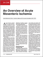
Arash Mirrahimi, MD, MSc; Charlotte Gallienne, MD; Hournaz Ghandehari, MD, FRCPC
Acute mesenteric ischemia (AMI) is a true surgical emergency and a rare life-threatening condition, accounting for 0.01% of hospital admissions, with extremely high mortality rates (up to 69%). Poor outcomes remain prevalent despite advances in both diagnostic and treatment options over the last two decades
Early diagnosis and management are particularly important given that the highest incidence of AMI occurs in the elderly population, who often have multiple comorbidities leading to a worse prognosis. Biphasic contrast enhanced multidetector computed tomography (MDCT) images have become the mainstay and standard of care for investigation and timely diagnosis of AMI.
As such, the importance of recognizing imaging features of AMI and timely communication of findings with the referring physicians is of utmost importance for diagnostic radiologists and always a worthwhile topic for review. We have therefore endeavored to provide a brief summary of the presentation of AMI, its causes, relevant anatomy, and most importantly, illustrated review of CT findings that delineate ischemic changes of the bowel and mesentery.
Available for SA-CME Credit. To receive SA–CME credit, you must complete the post exam and review the discussion and references provided with the exam results.
|
|
|
|
|
* Advanced MRI Safety Training For Healthcare Professionals (4th Edition): Level 2 MR Personnel
|
|
Released:
April 22, 2020
•Expires:
March 31, 2025
|
CE credits:
3.0
• Cost:
$50.00
|
|
Faculty:
Frank G. Shellock, PhD, FACC, FACR, FACSM
|
This program reviews fundamental MRI safety information and meets the annual training recommendations from the American College of Radiology. Importantly, MRI facilities must now comply with the revised requirements for diagnostic imaging from The Joint Commission and document that MRI technologists participate in ongoing education that includes annual training on safe MRI practices in the MRI environment. Notably, Advanced MRI Safety Training for Healthcare Professionals, Level 2 MR Personnel covers each MRI safety topic specified by The Joint Commission, as well as many additional subjects that will expand the knowledge-base of healthcare professionals involved with MRI technology.
With 35 years of experience in the field of MRI, the author of the best-selling textbook series, the Reference Manual for Magnetic Resonance Safety, Implants and Devices, and the creator of the internationally popular website, MRIsafety.com, Dr. Frank G. Shellock is uniquely qualified to present the information in this program.
Advanced MRI Safety Training for Healthcare Professionals (4th Edition), Level 2 MR Personnel is a 2 hour and 45 minute program that is divided into three different sections.
Educational Objectives
-
Understand the safety issues related to MRI.
-
Describe the bioeffects associated with the static magnetic field, time-varying magnetic fields, and radiofrequency fields.
-
Present guidelines that prevent projectile-related accidents.
-
Describe polices that avoid issues related to acoustic noise.
-
Review procedures that prevent burns associated with MRI.
-
Explain and demonstrate appropriate pre-MRI screening procedures.
-
Identify techniques to manage patients with claustrophobia, anxiety, or emotional distress.
-
Describe guidelines to handle medical emergencies in the MRI setting.
-
Understand the safety considerations for gadolinium-based contrast agents.
This is a Pay-To-View program. Purchase is required for full program access.
|
|
|
|
|
* The Online Course for MR Safety Officers (MRSO) and MR Medical Directors (MRMD)
|
|
Released:
September 29, 2022
•Expires:
September 30, 2025
|
CE credits:
10.0
• Cost:
$975.00
|
|
Faculty:
William Faulkner, B.S.,R.T.(R)(MR)(CT), FSMRT | Kristan Harrington, MBA, R.T.(R)(MR), ARRT, MRSO
|
Applied Radiology and William Faulkner & Associates are pleased to introduce “The Online Course for MR Safety Officers (MRSO) and MR Medical Directors (MRMD)”. This comprehensive program, focusing on MR Safety, covers many aspects relating to the safety practices of the Magnetic Resonance Imaging (MRI) environment. It is designed for those individuals who are either currently serving as their facility’s MRSO and/or MRMD, or those who are preparing to assume these responsibilities. The content of this course will be helpful for those preparing for the American Board of Magnetic Resonance Safety MRSO and MRMD examinations.
The content of The Online Course for MR Safety Officers and MR Medical Directors is based upon current FDA and ACR guidelines, including the ACR Guidance Document for MR Safe Practices, as well as those promulgated by industry regulatory bodies such as the International Electrotechnical Commission.
The complete online program has been approved for up to 10 AMA PRA Category 1 credits™ (CME) and 10 Category A ARRT continuing educational credits (CE).
Module 1: Basic MRI Physics Relevant to MRI Safety
Module 2: Static Field: Bioeffects and Access Control
Module 3: Gradient Magnetic Fields: Bioeffects and Safety
Module 4: Radio Frequency Field: Bioeffects and Safety
Module 5: Implants and Devices
Module 6: Gadolinium-Based MR Contrast Agents
Module 7: MR Safety Screening
Module 8: Managing Emergent Situations and Patient Considerations: Quench and Patient Anxiety & Patient Monitoring
The Online Course for MR Safety Officers (MRSO) and MR Medical Directors (MRMD) is not affiliated with, nor endorsed by the American Board of Magnetic Resonance Safety (ABMRS)
Terms and Conditions of Use: Click To View
This is a Pay-To-View program. Purchase is required for full program access.
|
|
|
|
|
Evidence-based Approach to Minimizing IV Contrast Extravasations
|
|
Released:
July 10, 2019
•Expires:
December 31, 2027
|
CE credits:
1.0
• Cost:
$0.00
|
|
Faculty:
Ryan K Lee, MD, MBA
|
IV contrast extravasation is a not uncommon complication in the performance IV contrast enhanced CT examinations. In this accredited Expert Forum Webcast, we will review the different complications related to IV contrast administration. This will be followed by analysis of different factors that contribute to IV contrast-extravasation and subsequent discussion of best practices that can reduce the incidence of IV contrast extravasation. A novel method which reduced contrast extravasation by over 50% will also be presented.
Following the presentation questions from the audience were addressed in a moderated Q&A session.
Learning Objectives
-
Discuss the different types of complications associated with IV contrast extravasation.
-
Describe the various factors that can contribute to IV contrast extravasation.
-
Apply best practices to reduce the incidence of IV contrast extravasation.
No special educational preparation is required for this CME/CE Activity!
Supported through an unrestricted educational grant from Bracco Diagnostics, Inc.
|
|
|
|