|
Multimodality breast imaging, including MR imaging
Gillian M. Newstead, MD, FACR
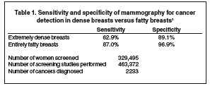
Dr. Newstead is a Professor of Radiology and the Clinical Director of Breast Imaging, the University of Chicago, Chicago, IL. Breast imaging has advanced significantly since the time when film-based mammography was the only imaging tool available for breast cancer diagnosis. Today, multimodality screening and diagnosis employing analog and digital mammography, ultrasound, and magnetic resonance (MR) imaging of the breast is a clinical reality. Breast cancer screening Mammography It is currently recommended that all women begin getting annual mammograms at the age of 40. Testing should begin sooner if the patient is at increased risk for breast cancer through a family history of premenopausal cancer, genetic disposition or through prior chest irradiation (eg, treatment for Hodgkins disease). Such high-risk patients should be referred to a high-risk assessment clinic that can provide the patient with all the necessary information and make a judgment as to whether or not additional screening methods, such as whole-breast ultrasound or screening MR, might be appropriate. Both analog and digital mammography screening have been found to be effective in detecting cancer, but this effectiveness can be limited by a variety of factors, including patient age and breast density. One recent study found that mammography has a sensitivity of 87.0% for detecting lesions in fatty breasts but that that number dropped to 62.9% in dense breasts (Table 1).1 Other factors, such as image quality and the experience and training of the interpreting radiologist, also play a role. Ultrasound screening According to general estimates, there are approximately 70 million U.S. women eligible for mammography today, 40 million of whom undergo mammography screening. Of those screened women, 90% are told they are healthy based on the mammographic study, but a small percentage —roughly 40,000—will actually have cancers that were missed at mammography. Ultrasound screening may be beneficial in detecting some of these mammographically occult cancers. Although there is no randomized controlled trial data to support the efficacy of ultrasound screening, the next American College of Radiology Imaging Network trial results (ACRIN 6666) should provide important additional information. Screening MR The detection rate of screening MR is significantly higher than that of ultrasound; however, the high cost per patient of MR is a concern. MR screening should be reserved for only those women who are at extremely high risk for breast cancer, such as those who are genetically predisposed or have at least a 20% to 25% lifetime risk of developing breast cancer. Women who carry the BRCA 1 or 2 gene have up to an 85% lifetime risk of developing breast cancer. These women also tend to have an earlier onset of disease and a higher prevalence for bilateral disease. These are the types of patients who should be considered candidates for MR breast cancer screening and should be evaluated in a high-risk– assessment clinic. Despite the high per-study cost of MR, if this screening method is reserved for only extremely high-risk women, its high yield will result in a cost per cancer found that is similar to the cost of finding a cancer in an average-risk woman with mammography. The yield is so much greater in the subset of patients who are at very high risk that the cost is about the same. When developing an MR screening program, it is important that the MR screening population be clearly defined. Once defined, it is important to monitor the program’s protocol to ensure that the guidelines are followed and that only the designated patients are included. Also, it is important to remember that the lesions that are found only with MR are most likely to be occult mammographically and ultrasonographically. Therefore, should a biopsy be required, it will need to be performed with MR guidance.
A recent study compared the sensitivity and specificity of clinical breast examination (CBE), MR imaging, and mammography for invasive tumor detection in women at high risk for breast cancer (Table 2).2 Over 4 years, nearly 2000 women were screened, 358 of whom had germ-line mutations. The participants underwent CBE every 6 months and mammography and MR breast examination annually. With a median follow-up of 2.9 years, 45 breast cancers were found: 39 invasive cancers and 6 cases of ductal carcinoma in situ (DCIS). The overall detection rate was 9.5 per 1000 for all participants and 26.5 per 1000 in those with the genetic mutation. The sensitivity for CBE was found to be 18% (98% specificity), for mammography it was 33% (95% specificity), and for MR imaging it was 80% (90% specificity). The authors also found that patients who carried the gene mutation also tended to have larger lesions upon detection. In this study, 43% of the patients in the MR surveillance group had lesions <10 mm, compared with only 12% or 14% of those in the 2 control groups. In addition, women screened with MR were less likely to have positive lymph nodes at diagnosis. In subsequent rounds, the more favorable prognosis for cancers detected in the group that was screened with MR was maintained.3 Breast MR imaging How does MR imaging of the breast compare with mammography? With mammography screening, typically only 2 views of the breast are obtained. With the dynamic capabilities of MR imaging, the radiologist can see throughout the entire breast in a range of projections. Unlike X-ray technology, MR works well in women with dense breast tissue, in women with scar tissue from prior surgery, and in women with breast implants. With current MR imaging techniques, the calcifications commonly associated with noninvasive breast cancer (DCIS) are typically not seen. Rather, the radiologist looks for the angiogenic effect of the lesion. In addition, unlike mammographic examinations, MR imaging requires contrast in- jection and a longer examination time. The important advantage of a dy-namic contrast-enhanced MR is that virtually all cancers enhance following contrast injection. This means that nearly all cancers will be visible on MR. MR imaging also has a very high negative predictability for invasive cancer. Therefore, if a patient with a suspicious clinical or other imaging finding has a negative breast MR finding with no enhancement, it is very unlikely that the patient has an invasive breast cancer. This can be particularly important among patients whose imaging may be complex because of prior treatment or surgery. Tumor angiogenesis makes cancer highly visible on contrast-enhanced MR. The newly formed vessels tend be of poor quality and leak contrast around the tumor. With gadolinium injection, the alterations in the vasculature associated with cancer are clearly visible, often making it easy to distinguish malignant tumors from benign findings if the contrast study is performed properly. The degree of enhancement will depend on the histology of the underlying lesion, which also helps distinguish malignant lesions from benign ones. MR scanners For MR imaging of the breast, a dedicated breast coil is needed. The patient lies prone, and the breasts are suspended in the wells and immobilized slightly to avoid motion. Markers are placed on the nipples and any areas with scars, similar to what is done for mammography. Early breast coils had only 2 or 4 channels. Current systems have 7- and 8-channel coils, and some manufacturers are currently developing 13- to 16-channel coils. The advent of parallel imaging technology has dramatically improved image quality when used with multichannel breast coils. The challenges in breast MR include the issues of spatial and temporal resolution. Spatial resolution is essential in order to assess the morphology of lesions. All breast specialists are familiar with the process of assessing morphology on mammography and ultrasound. Translating those skills to MR imaging is very straightforward. With MR, however, the issue of temporal resolution is added. Good temporal resolution is needed to determine the kinetic functions of lesions. Uniform fat suppression to enhance conspicuity is also important. The 3-dimensional (3D) imaging capability, the fourth dimension of temporal resolution, and the exquisite soft tissue contrast that are possible with new MR technique, provide vast improvements in image quality. Temporal resolution allows radiologists to assess how quickly a lesion takes up contrast. In the initial phase of the examination, contrast wash-in is assessed. In the later phase, wash-out is assessed. Benign lesions tend to enhance slowly and exhibit little wash-out, while malignant lesions tend to enhance and wash out quickly. Many radiologists believe the first images should be obtained within the first 1 to 2 minutes following contrast injection. My personal opinion is that the closer to 1 minute, the better. The optimal time may vary somewhat, depending on the magnet as well as the software and hardware being used. In our practice, the most common indication for breast MR is evaluation of an index cancer or the presence of a highly suspicious lesion pre-biopsy. Approximately 20% of our patients who undergo screening MR do so because they are found to be at super high risk for breast cancer or because they are extremely concerned and their physicians ordered a screening MR. The remaining patients tend to be previously treated cancer patients who undergo breast MR for assessment of residual cancer questions, posttreatment recurrence, or any of a variety of problem-solving issues. Can breast MR improve the assessment of tumor size, margins, numbers, and locations of cancers in patients with newly diagnosed breast cancer? The answer to that question is unequivocally yes. We find additional cancers in our practice up to 30% of the time. Most of the lesions are in the same quadrant, but some are in different quadrants or even in the other breast. The MR findings alter therapy for roughly 25% of our patients overall. The big question is whether breast MRI affects survival. Are these cancers that we are finding actually going to be clinically important? I think the answer is yes. Many of these findings are additional invasive cancers in either the ipsilateral or contralateral breast that, if not diagnosed and treated appropriately, will present as either a recurrent tumor or a new tumor at a later date. MR and DCIS Initially it was thought that MR was not very useful in detecting DCIS. This may have been a consequence of the fact that DCIS manifests differently on MR than it does on mammography. On MR, DCIS typically presents as a nonmass enhancement (Figure 1) rather than as the calcifications commonly seen on a mammogram. The angiogenic effects of DCIS are generally visible on MR. Segmental nonmass enhancement, enhancement of the ducts, and linear enhancement are commonly seen. In some cases, DCIS can present as a mass, but this is less common. In our practice, we often find noncalcified, mammographically occult DCIS using MR. With DCIS, it is often difficult to accurately size the cancer; therefore, surgeons like to take wider margins when employing breast-conserving therapy. With MR, when we find noncalcified DCIS, we have to determine how we are going to localize it for the surgeon. Often, we re-image the patient and take magnification views with mammography, to see if we can retrospectively find the lesion, and at times we cannot. Therefore, in order to excise the lesion, we must do a segmental excision using a bracketed MR-guided needle localization procedure. MR is also useful for assessing extramammary nodal disease and for checking for tumor invasion into the chest wall. Because of the 3D format of MR, the posterior tissues can be clearly visualized and it is possible to determine if a posterior lesion has invaded the pectoral muscle. If enhancement of the pectoral muscle is seen, chest wall invasion is likely. It is also possible to visualize axillary lymph nodes with MR to determine if they are morphologically abnormal. In the future, lymphotropic nanoparticle imaging may provide improved sensitivity and specificity for microscopic nodal involvement. At present, we use ultrasound for assessment of nodal disease and core biopsy to document metastases if the nodes are morphologically abnormal. Effect on treatment At the University of Chicago, we followed 140 consecutive patients with noninflammatory cancer who underwent MR breast examination. In 40 (28.6%) of these patients, additional cancers were found on the MR study. Of those, 26 were found in the same quadrant as the initial cancer, 11 were in a different quadrant, and 3 were found in the contralateral breast. These findings altered therapy in 31 patients: 20 had larger lumpectomies than had been initially planned, 8 were determined to require mastectomy, and 3 were changed to neo-adjuvant treatment.4 With MR, additional cancer is found in approximately 30% of patients. This means that in roughly 70% of patients we can be confident that the cancer is confined to the extent determined prior to the MR study. That is important information when considering focal radiation treatment. MR imaging has a positive and reinforcing effect in cases in which the disease is truly unifocal. For these patients, alternative therapies, such as ablation procedures and more focal radiation therapy, may be options.
Other indications MR can also be used to evaluate a patient for residual cancer. Typically following surgery, a seroma cavity will be visible; enhancement of the seroma rim, typically 2 to 3 mm in thickness, is usually seen. Patients with positive margins should be imaged as soon as they can tolerate lying in the magnet. In these patients, mammography is not going to be useful, except perhaps in cases of DCIS in which the clinician is looking for residual calcifications. Noncalcified lesions will not be visible on a mammogram because of the presence of the large seroma cavity. In addition, the inability to obtain adequate compression in a postoperative patient will further limit mammography’s usefulness in these patients. MR can also be used to locate a small primary lesion in patients who present with palpable nodes or to assess for recurrent tumors. Many of these “recurrent tumors” may actually be tumors that were present but not initially detected or treated. It can also be used to assess patients who are difficult to image with mammography and/or ultrasound because of other factors, such as deformity, scarring, and postsurgical changes to the breast tissue. For such patients, it can often be much easier to make a diagnosis with MR imaging. Postsurgical follow-up of certain patients may also be an indication for MR imaging, particularly if the initial cancer was mammographically occult. MR can also be used to measure response to tumor therapy. A study by Warren et al5 showed that sensitivity and specificity of MR were superior to conventional imaging for monitoring response to chemotherapy. Conclusion In our institution, MR imaging of newly diagnosed breast cancer patients has become routine practice. The surgical, medical, and radiation oncologists now depend on the information provided by MR to assist them in formulating de-finitive treatment measures. MR screening of high-risk women is also an important role for breast MR imaging. It is probable that higher field-strength magnets, innovative protocols, and new contrast agents will continue to improve and expand the applications for breast MR imaging in the future.
REFERENCES |

